Copyright
©The Author(s) 2023.
World J Orthop. Oct 18, 2023; 14(10): 733-740
Published online Oct 18, 2023. doi: 10.5312/wjo.v14.i10.733
Published online Oct 18, 2023. doi: 10.5312/wjo.v14.i10.733
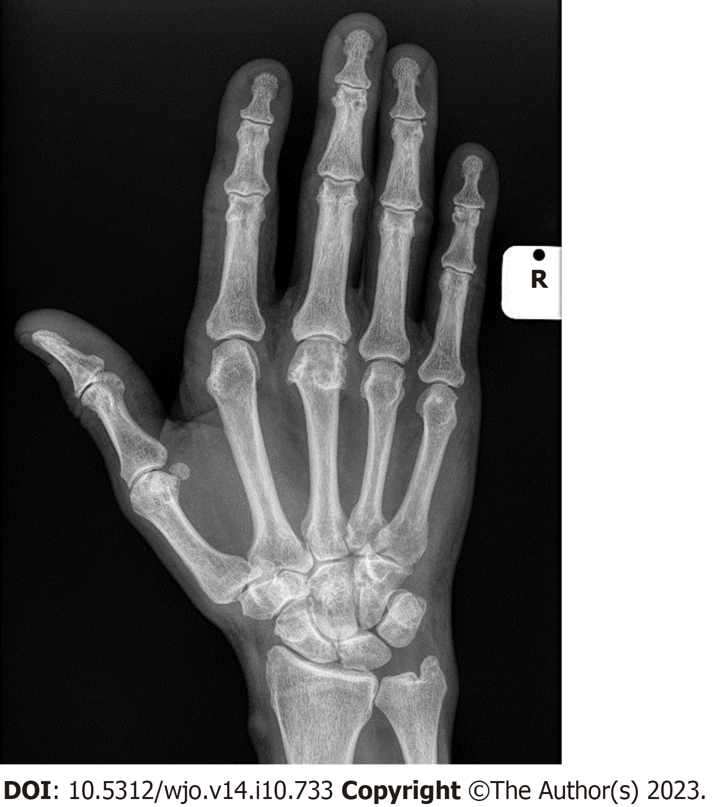
Figure 1 X-ray of a hand with crepitus.
Osteoarthritis of the middle finger metacarpophalangeal joint.
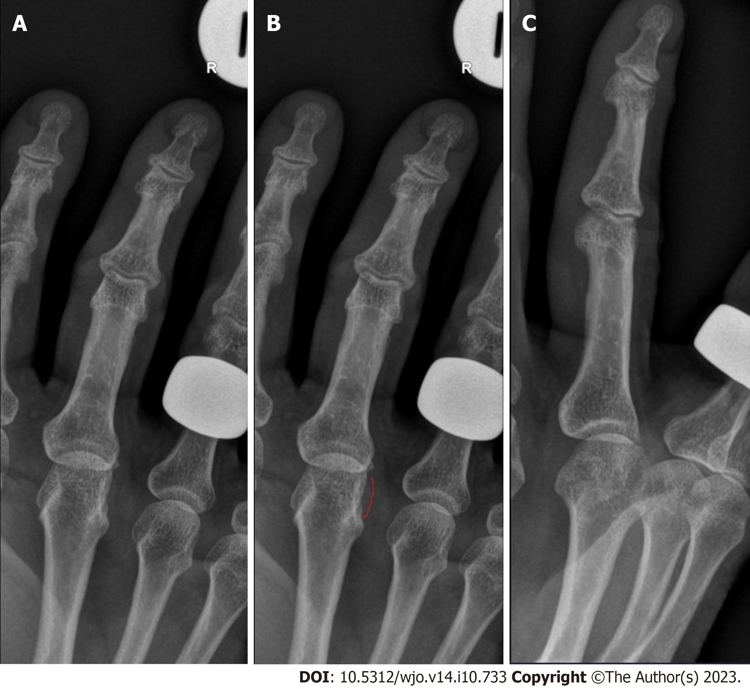
Figure 2 X-ray of a hand with locking.
A: X-rays of locked metacarpophalangeal joint (MCPJ); B: X-rays with ulnar osteophyte marked; C: Oblique view of locked MCPJ.
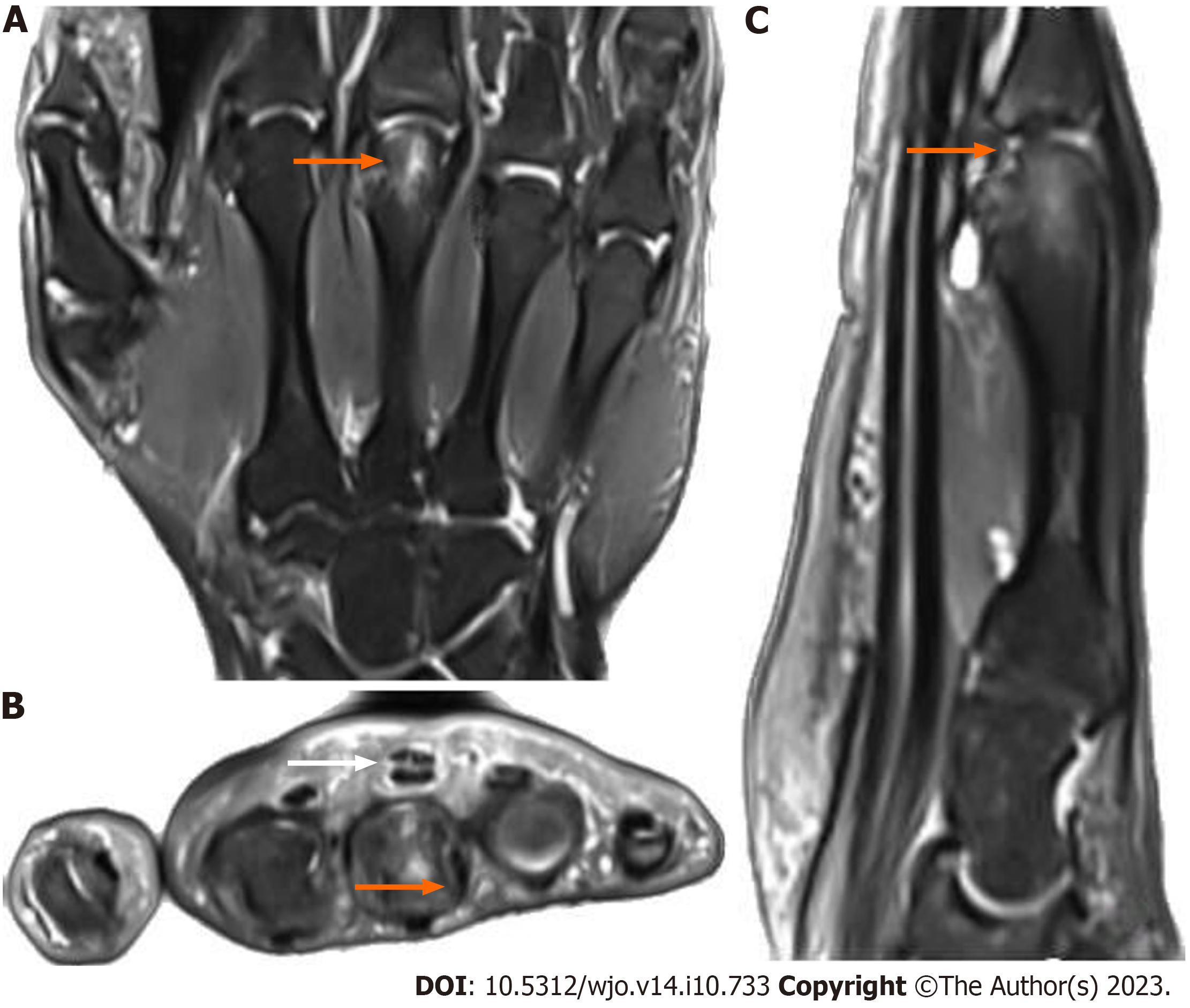
Figure 3 Magnetic resonance imaging of a hand with pain and symptoms.
A: Edema in the third metacarpal head (orange arrow) shown on magnetic resonance imaging (MRI) coronal view T2; B: Edema in the third metacarpal head (orange arrow) and flexor tendons with minimal effusion (white arrow) shown on magnetic resonance imaging transverse view; C: Edema in the third metacarpal head with effusion and joint surface involvement (orange arrow) shown on MRI sagittal view T2.
- Citation: Jordaan PW, Klumpp R, Zeppieri M. Triggering, clicking, locking and crepitus of the finger: A comprehensive overview. World J Orthop 2023; 14(10): 733-740
- URL: https://www.wjgnet.com/2218-5836/full/v14/i10/733.htm
- DOI: https://dx.doi.org/10.5312/wjo.v14.i10.733


