Copyright
©The Author(s) 2022.
World J Orthop. Sep 18, 2022; 13(9): 870-875
Published online Sep 18, 2022. doi: 10.5312/wjo.v13.i9.870
Published online Sep 18, 2022. doi: 10.5312/wjo.v13.i9.870
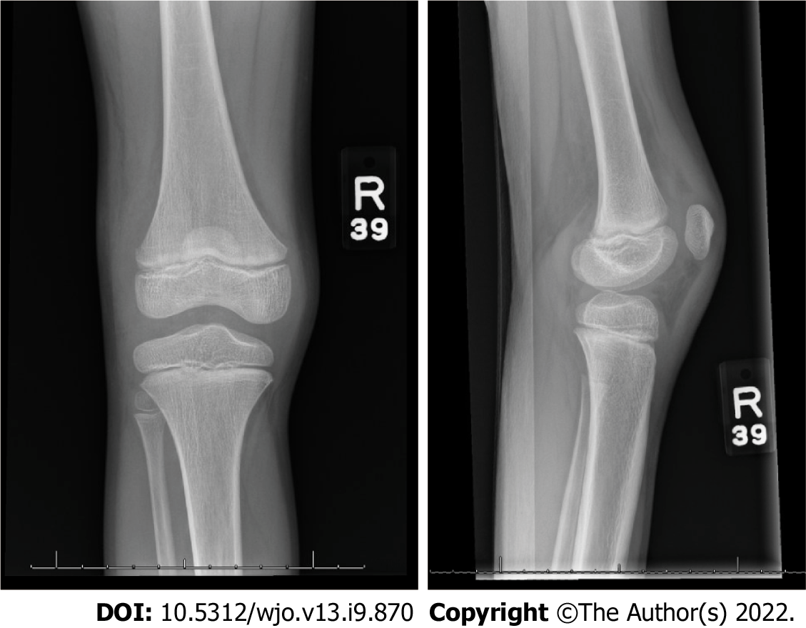
Figure 1 Anteroposterior (left) and lateral (right) radiographs of the right knee demonstrating evidence of a joint effusion within the suprapatellar recess and Hoffa’s fat pad.
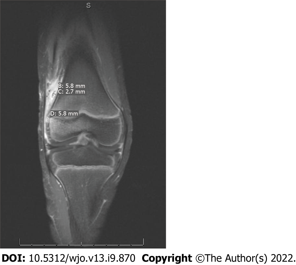
Figure 2 Magnetic resonance imaging with intravenous contrast of the right knee demonstrating a small enhancing cortical defect along the lateral border of the lateral femoral condyle, measuring approximately 6 mm, suggestive of osteomyelitis.
There is a collection within the inflammatory changes of the vastus lateralis demonstrating rim enhancement measuring approximately 0.6 cm × 0.2 cm representing tiny abscess formation.
- Citation: Pavlis W, Constantinescu DS, Murgai R, Barnhill S, Black B. Calcium pyrophosphate dihydrate crystals in a 9-year-old with osteomyelitis of the knee: A case report. World J Orthop 2022; 13(9): 870-875
- URL: https://www.wjgnet.com/2218-5836/full/v13/i9/870.htm
- DOI: https://dx.doi.org/10.5312/wjo.v13.i9.870


