Copyright
©The Author(s) 2022.
World J Orthop. Aug 18, 2022; 13(8): 679-692
Published online Aug 18, 2022. doi: 10.5312/wjo.v13.i8.679
Published online Aug 18, 2022. doi: 10.5312/wjo.v13.i8.679
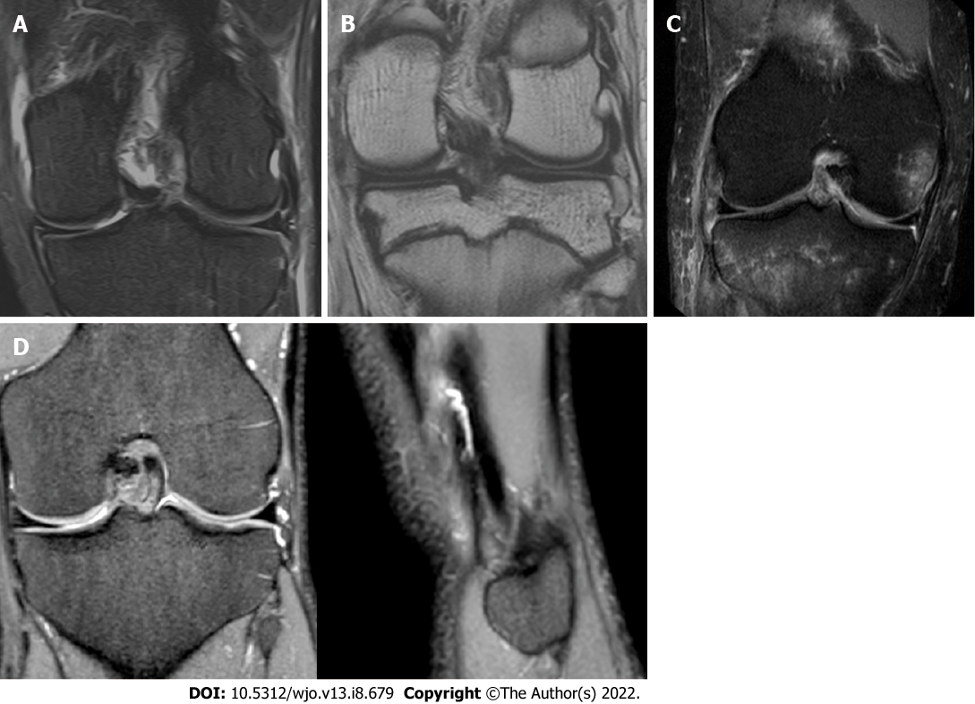
Figure 1 Magnetic resonance imaging.
A: Coronal T2 magnetic resonance imaging (MRI) showing a partial anterolateral ligament (ALL) lesion; B: Coronal T1 MRI showing a partial ALL lesion; C: Coronal STIR MRI showing a complete ALL lesion; D: Coronal and Sagittal T2 MRI showing an intact ALL.
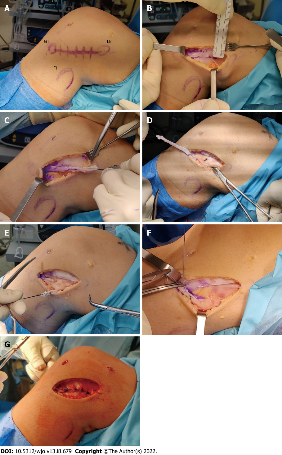
Figure 2 Cocker Arnold Mod Technique.
A: Bony landmarks on the anterolateral aspect of the knee: Gerdy’s tubercle, lateral epicondyle, fibular head; B: An iliotibial band (ITB) strip 1cm-wide, 8 cm-long is prepared; C: ITB strip harvesting maintaining the distal insertion; D: Identification of the fibular collateral ligament (FCL) and blunt dissection; E: The graft is passed under the FCL and reflected to itself; F: The graft is sutured to Gerdy’s tubercle with knee in extension and neutral rotation; G: The ITB is sutured in a standard fashion.
- Citation: Sabatini L, Capella M, Vezza D, Barberis L, Camazzola D, Risitano S, Drocco L, Massè A. Anterolateral complex of the knee: State of the art. World J Orthop 2022; 13(8): 679-692
- URL: https://www.wjgnet.com/2218-5836/full/v13/i8/679.htm
- DOI: https://dx.doi.org/10.5312/wjo.v13.i8.679


