Copyright
©The Author(s) 2022.
World J Orthop. May 18, 2022; 13(5): 427-443
Published online May 18, 2022. doi: 10.5312/wjo.v13.i5.427
Published online May 18, 2022. doi: 10.5312/wjo.v13.i5.427
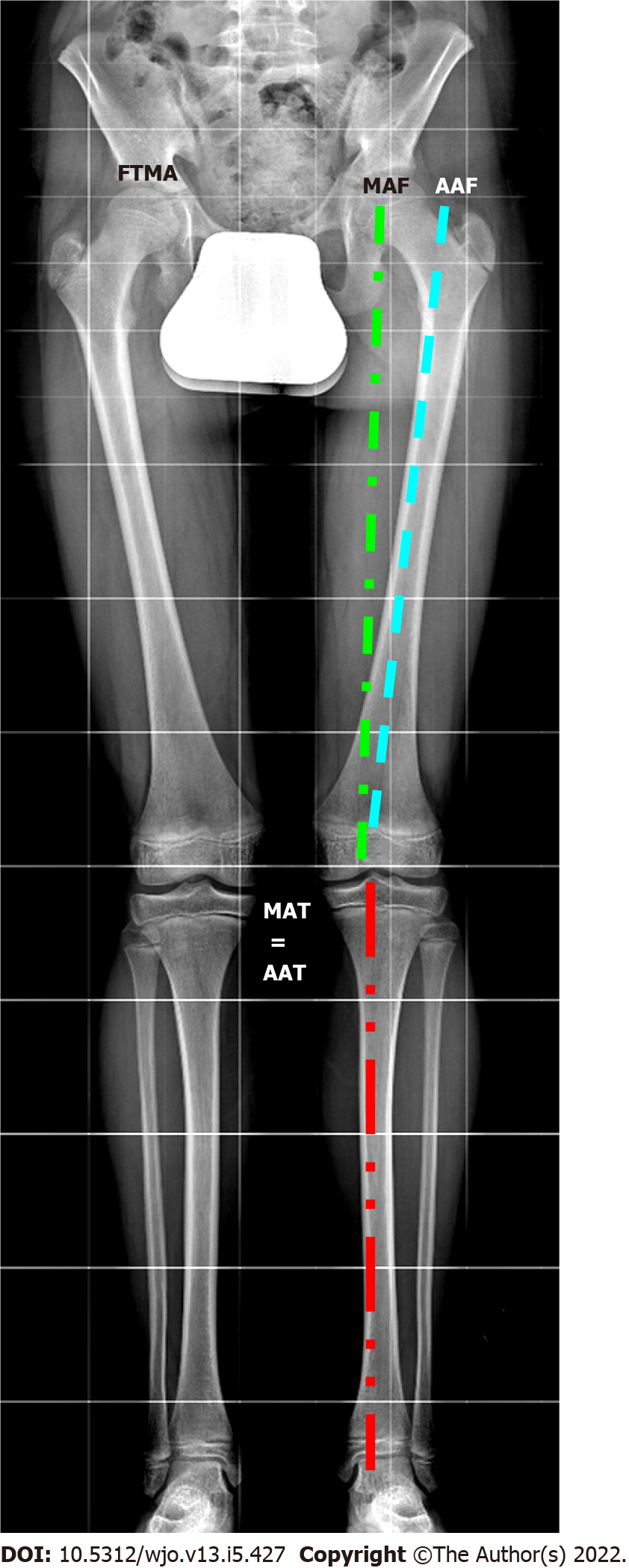
Figure 1 Lower limb axes evaluated in long standing X-ray.
FTMA: Femorotibial mechanical axis; MAF: Mechanical axis of the femur; AAF: Anatomical axis of the femur; MAT: Mechanical axis of the tibia; AAT: Anatomical axis of the tibia.
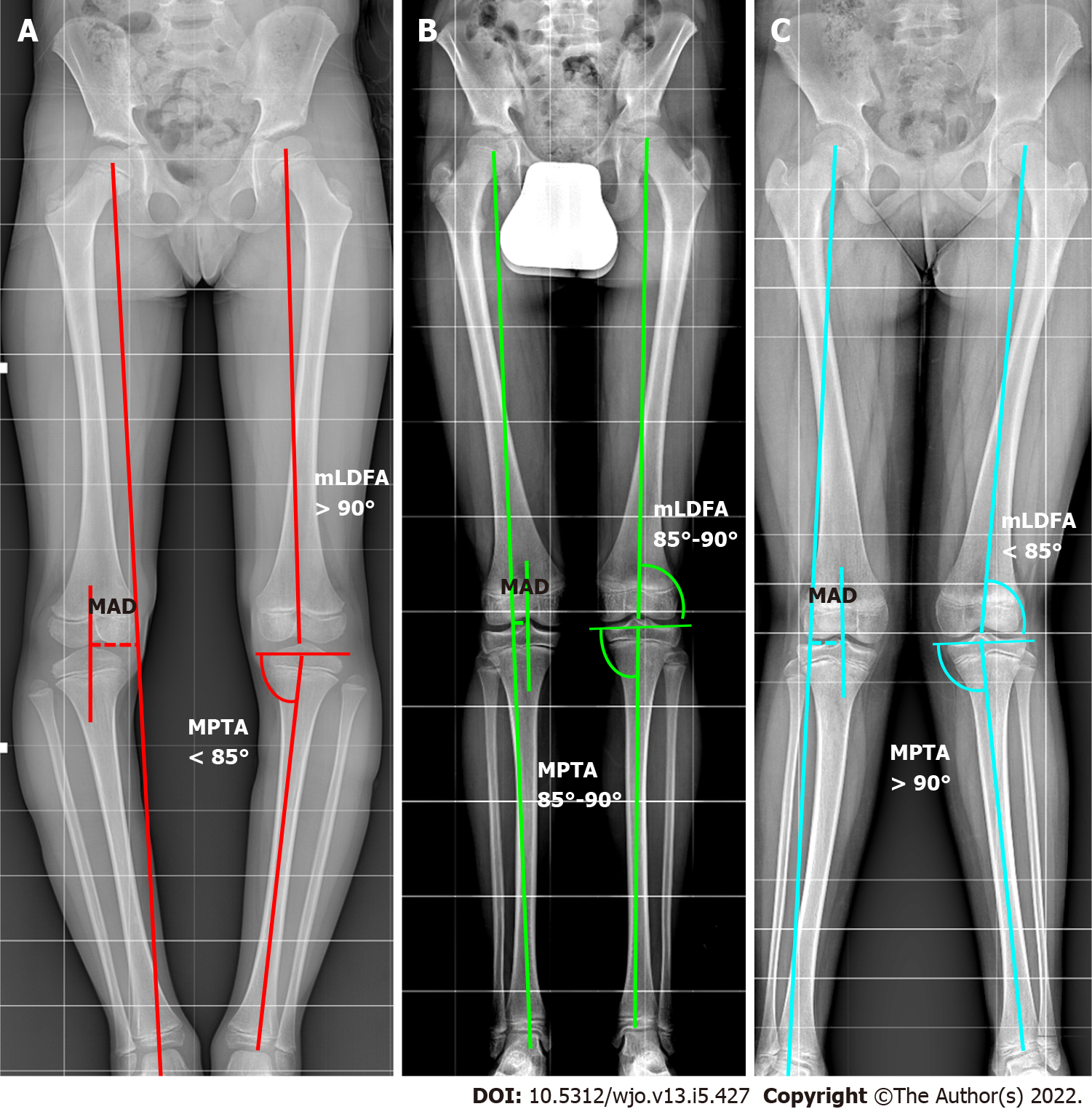
Figure 2 The malalignment test.
The mechanical axis (MA) is traced from the center of the femoral head to the center of the ankle. The metaphyseal-diaphyseal angle (MAD) is calculated in millimeters (dotted lines in the image traced from center of knee and MA). If the MAD exceeds the threshold of normality, it is necessary to find the source of the deformity. The mechanical lateral distal femur angle. Medial proximal tibial angle are evaluated. A: Varus; B: Normal; C: Valgus. MAD: Metaphyseal-diaphyseal angle; MPTA: Medial proximal tibial angle; mLDFA: Mechanical lateral distal femur angle.
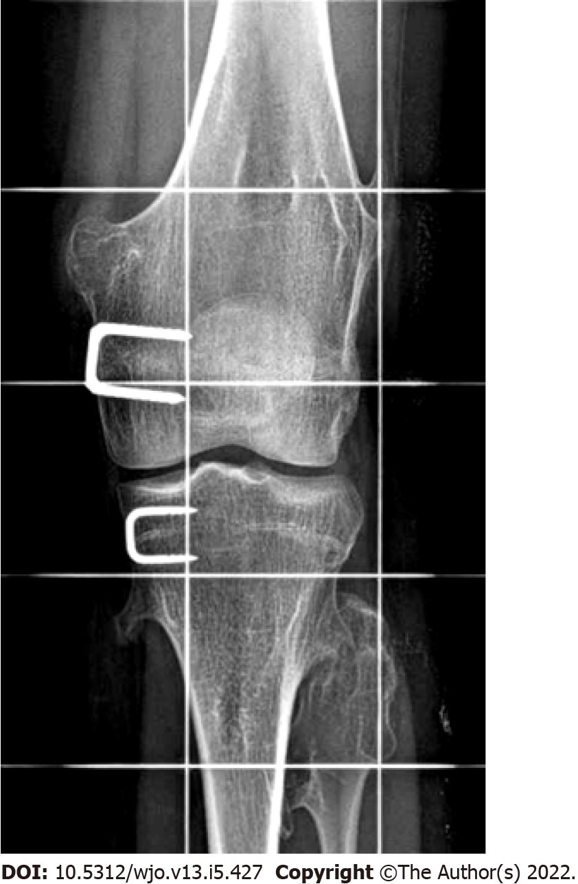
Figure 3 Staple hemiepiphysiodesis.
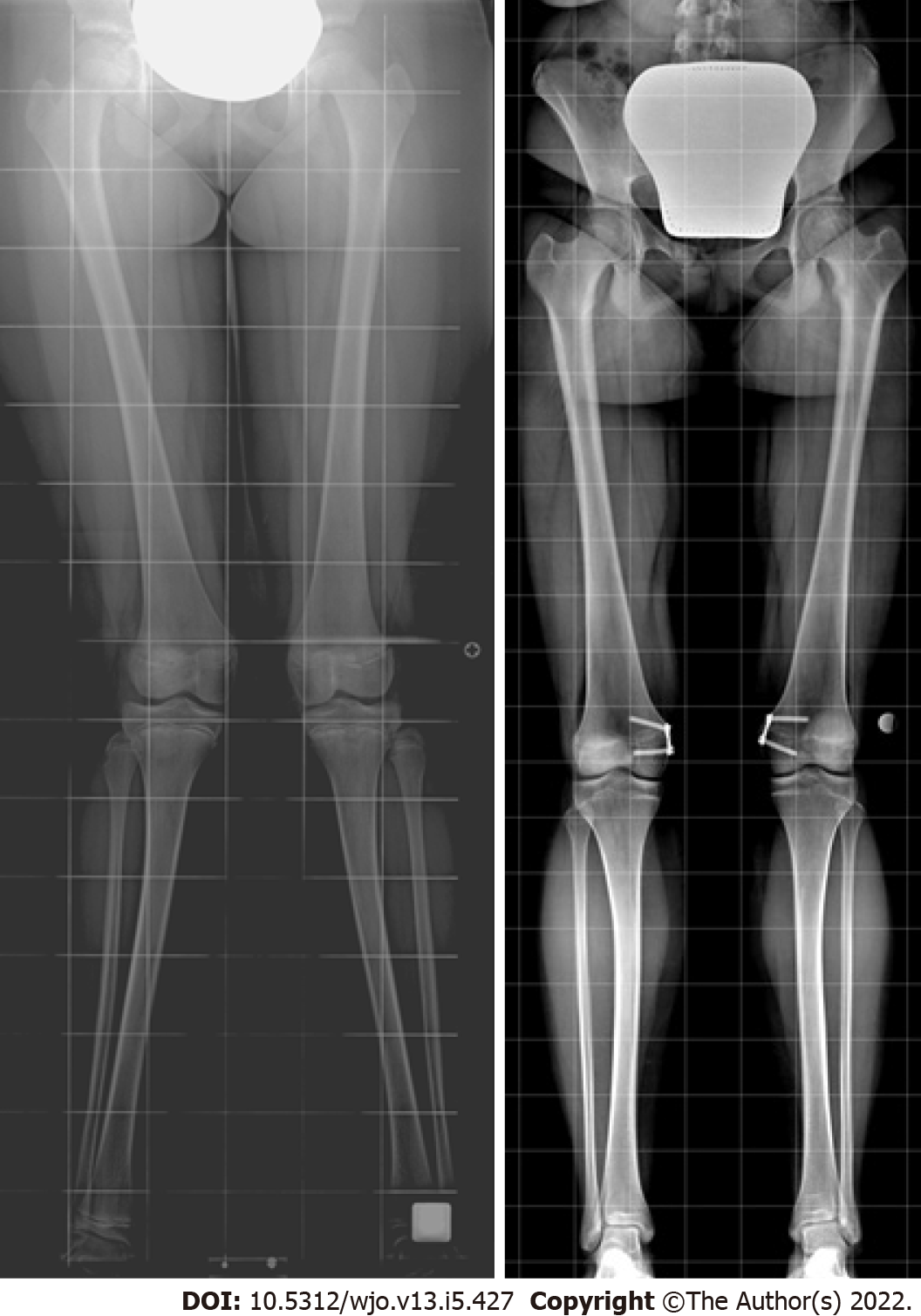
Figure 4 Tension band plate hemiepiphysiodesis for correcting genu valgum deformity.
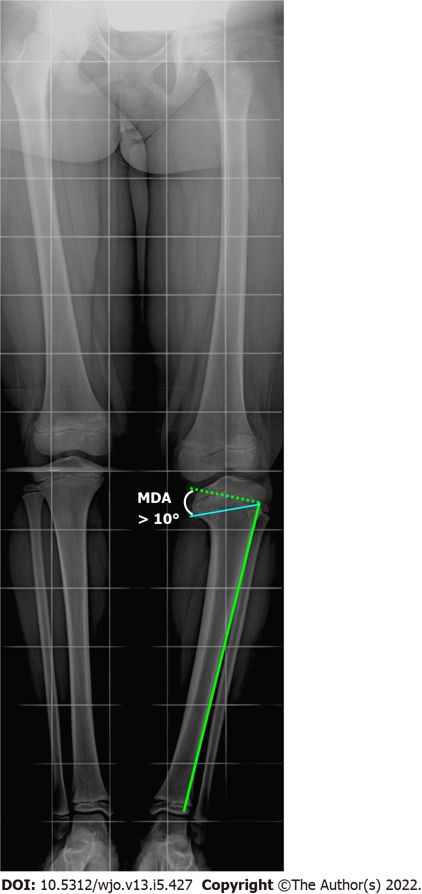
Figure 5 Radiographic measurement of Drennan’s metaphyseal-diaphyseal angle.
The metaphyseal-diaphyseal angle (MDA) is measured from a perpendicular line to the tibial diaphyseal axis and a line passing through the axial plane of the proximal tibial metaphysis. An MDA > 10 degrees associated with a tibiofemoral angle > 20 degrees indicates a toddler at risk. MDA: Metaphyseal-diaphyseal angle.
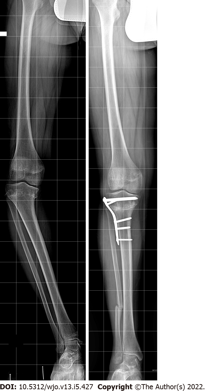
Figure 6 High tibial osteotomy for genu varum correction.
- Citation: Coppa V, Marinelli M, Procaccini R, Falcioni D, Farinelli L, Gigante A. Coronal plane deformity around the knee in the skeletally immature population: A review of principles of evaluation and treatment. World J Orthop 2022; 13(5): 427-443
- URL: https://www.wjgnet.com/2218-5836/full/v13/i5/427.htm
- DOI: https://dx.doi.org/10.5312/wjo.v13.i5.427


