Copyright
©The Author(s) 2022.
World J Orthop. Apr 18, 2022; 13(4): 400-407
Published online Apr 18, 2022. doi: 10.5312/wjo.v13.i4.400
Published online Apr 18, 2022. doi: 10.5312/wjo.v13.i4.400
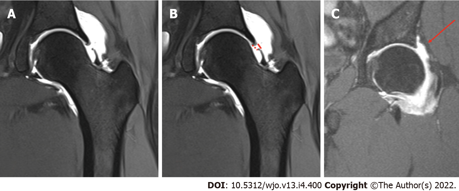
Figure 1 Example of capsular defect and intact capsule on magnetic resonance imaging-arthrography.
A: example of a capsular defect on magnetic resonance imaging (MRI)-arthrography with extracapsular contrast leakage to the adjacent soft-tissue; B: Gap length measurement; solid line: gap length muscular side. Dotted line: Gap length acetabular side; C: Example of an intact capsule on MRI-arthrography (Arrow). There is no contrast leakage to the adjacent soft-tissue.
- Citation: Bech NH, van Dijk LA, de Waard S, Vuurberg G, Sierevelt IN, Kerkhoffs GM, Haverkamp D. Integrity of the hip capsule measured with magnetic resonance imaging after capsular repair or unrepaired capsulotomy in hip arthroscopy. World J Orthop 2022; 13(4): 400-407
- URL: https://www.wjgnet.com/2218-5836/full/v13/i4/400.htm
- DOI: https://dx.doi.org/10.5312/wjo.v13.i4.400


