Copyright
©The Author(s) 2022.
World J Orthop. Apr 18, 2022; 13(4): 381-387
Published online Apr 18, 2022. doi: 10.5312/wjo.v13.i4.381
Published online Apr 18, 2022. doi: 10.5312/wjo.v13.i4.381
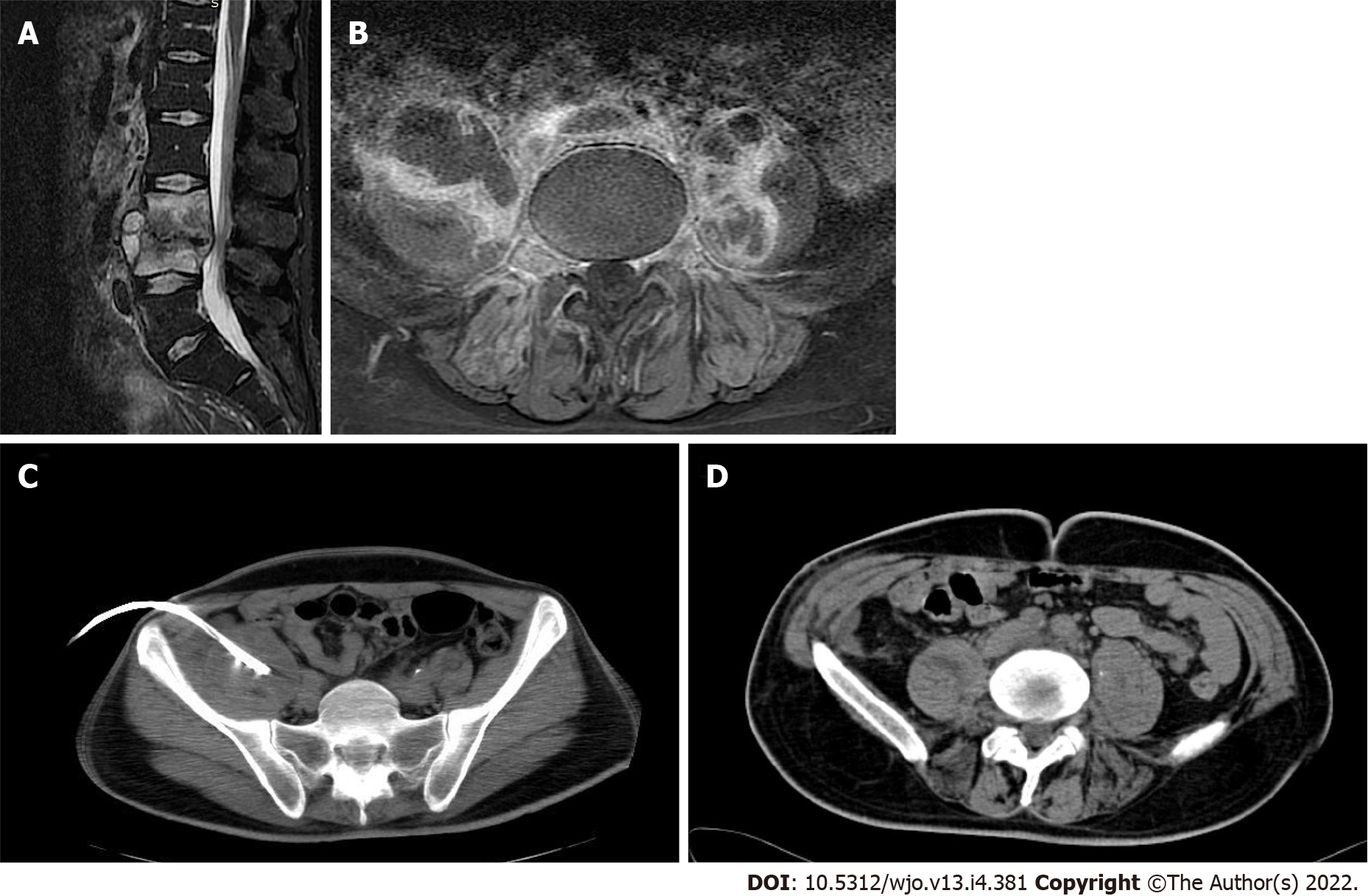
Figure 1 A 35-year-old patient with a history of intravenous drug use presenting with severe low back pain.
A: T2 weighted image sagittal image reveals a high-intensity signal of the L3-L4 vertebrae, disk involvement, and paravertebral fluid collections; B: T1 weighted image axial image with contrast enhancement reveals bilateral iliopsoas abscesses; C: The corresponding computed tomography image with the pigtail catheter inserted in the right iliopsoas abscess; D: The computed tomography image after the catheter removal revealed complete resolution of the abscess.
- Citation: Fesatidou V, Petsatodis E, Kitridis D, Givissis P, Samoladas E. Minimally invasive outpatient management of iliopsoas muscle abscess in complicated spondylodiscitis. World J Orthop 2022; 13(4): 381-387
- URL: https://www.wjgnet.com/2218-5836/full/v13/i4/381.htm
- DOI: https://dx.doi.org/10.5312/wjo.v13.i4.381


