Copyright
©The Author(s) 2021.
World J Orthop. Sep 18, 2021; 12(9): 710-719
Published online Sep 18, 2021. doi: 10.5312/wjo.v12.i9.710
Published online Sep 18, 2021. doi: 10.5312/wjo.v12.i9.710
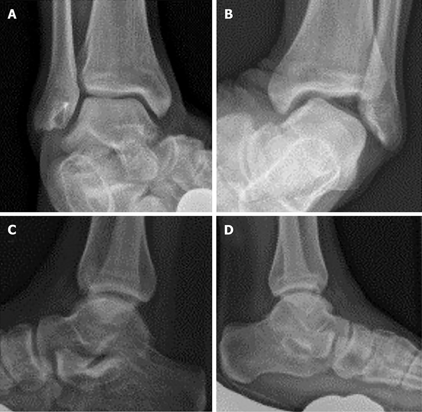
Figure 1 Stress radiographs.
A: An example of a stable ankle which has previously undergone a modified Broström procedure is depicted; B: The contralateral ankle demonstrating instability on the talar tilt examination is depicted; C and D: The corresponding anterior drawer stress radiographs.
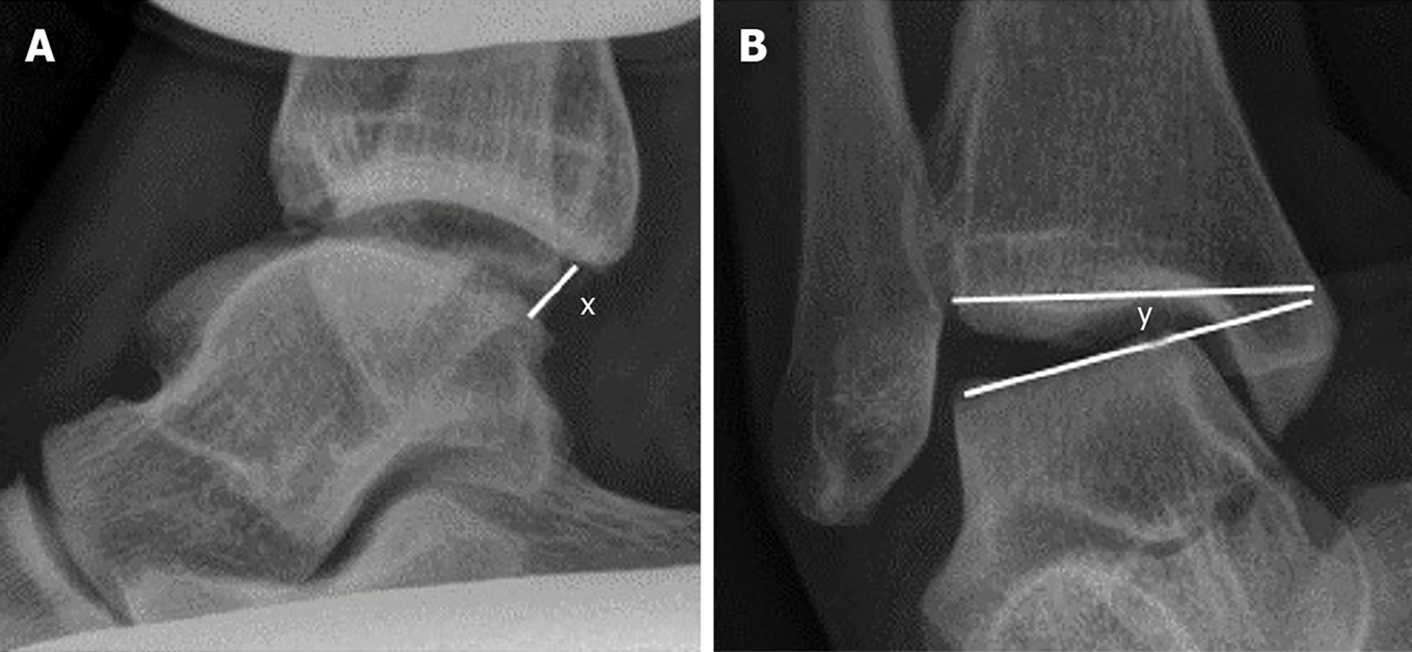
Figure 2 The measurements for stress radiographs are depicted here.
A: The anterior drawer distance is obtained by drawing a line from the most posterior aspect of the distal tibial plafond to a point on the talus that is perpendicular to the articular surface of the talus. The distance ‘x’ is measured in millimeters; B: The anterior drawer is depicted. In order to measure the anterior drawer one line is drawn along the distal articular surface of the tibia and a second line is drawn along the proximal articular surface of the talus. The angle ‘y’ formed by the intersection of these two lines is the talar tilt angle and is measured in degrees.
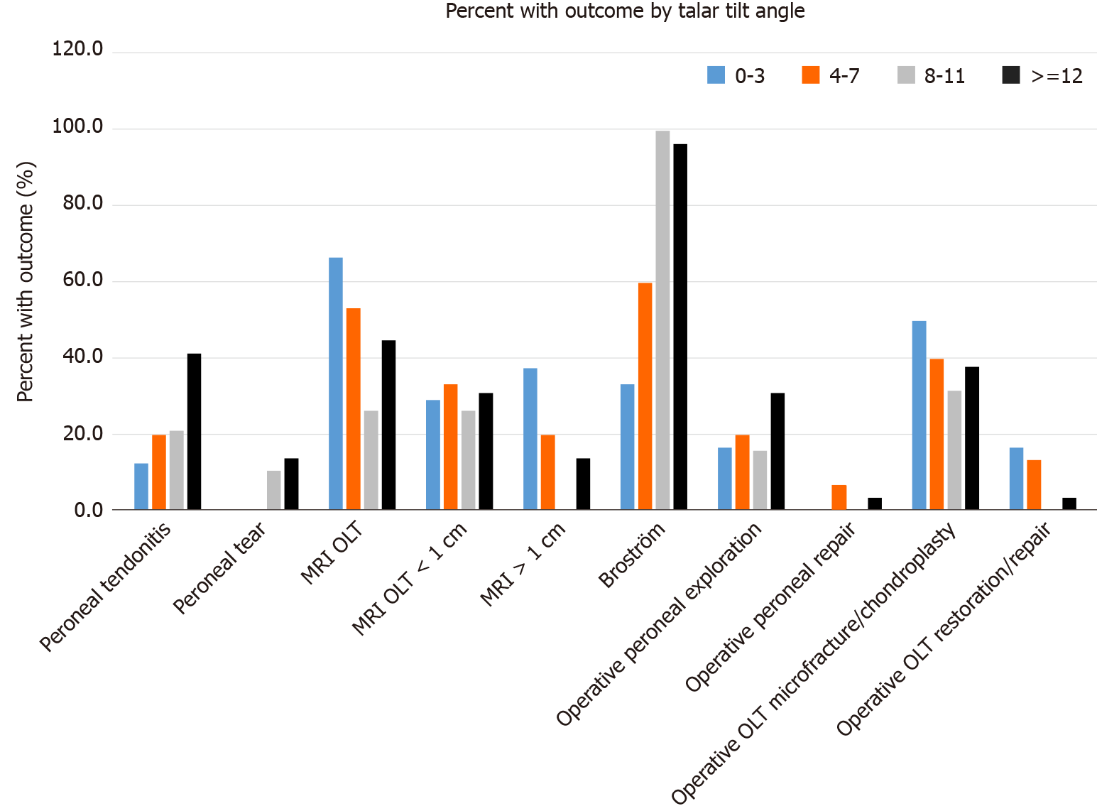
Figure 3 The presence of an associated condition is listed next to the number of ankles that have that condition when organized according to the degree of instability as measured on the talar tilt image.
MRI: Magnetic resonance imaging; OLT: Osteochondral lesions of the talus.
- Citation: Sy JW, Lopez AJ, Lausé GE, Deal JB, Lustik MB, Ryan PM. Correlation of stress radiographs to injuries associated with lateral ankle instability. World J Orthop 2021; 12(9): 710-719
- URL: https://www.wjgnet.com/2218-5836/full/v12/i9/710.htm
- DOI: https://dx.doi.org/10.5312/wjo.v12.i9.710


