Copyright
©The Author(s) 2021.
World J Orthop. Sep 18, 2021; 12(9): 660-671
Published online Sep 18, 2021. doi: 10.5312/wjo.v12.i9.660
Published online Sep 18, 2021. doi: 10.5312/wjo.v12.i9.660
Figure 1 Plain radiographs before and after atypical bisphosphonate associated femoral fracture fixation.
A: Before atypical bisphosphonate associated femoral fracture fixation; B: After atypical bisphosphonate associated femoral fracture fixation.
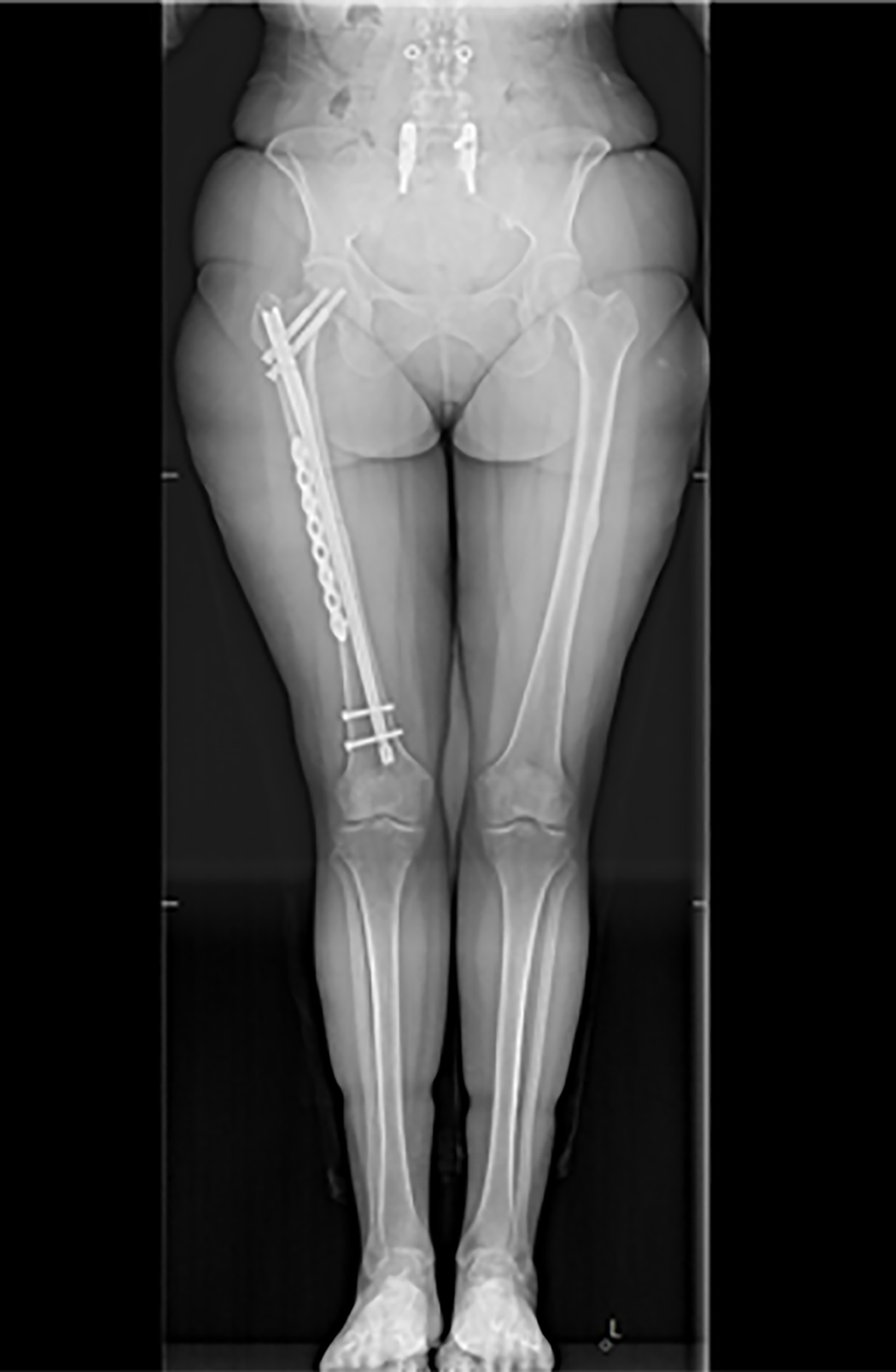
Figure 2 Plain radiograph illustrating fixation of an atypical bisphosphonate associated fracture and beaking on the contralateral limb at the same level.
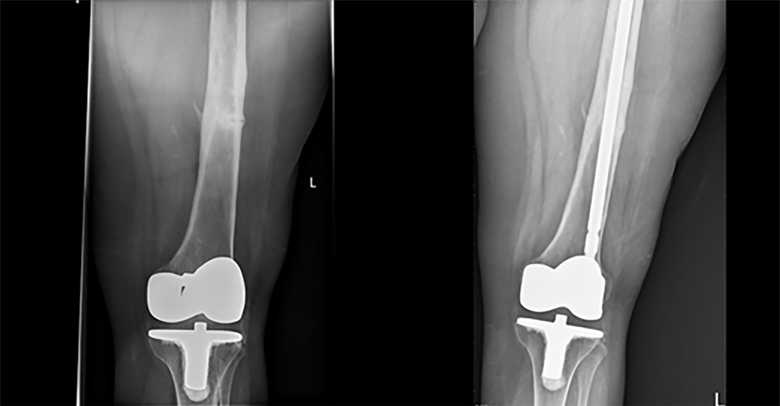
Figure 3 Plain radiographs of the “dreaded lucent line” and distal unlocked intramedullary stabilisation to minimise the stress riser around a knee replacement.
- Citation: Rudran B, Super J, Jandoo R, Babu V, Nathan S, Ibrahim E, Wiik AV. Current concepts in the management of bisphosphonate associated atypical femoral fractures. World J Orthop 2021; 12(9): 660-671
- URL: https://www.wjgnet.com/2218-5836/full/v12/i9/660.htm
- DOI: https://dx.doi.org/10.5312/wjo.v12.i9.660


