Copyright
©The Author(s) 2021.
World J Orthop. Aug 18, 2021; 12(8): 534-547
Published online Aug 18, 2021. doi: 10.5312/wjo.v12.i8.534
Published online Aug 18, 2021. doi: 10.5312/wjo.v12.i8.534
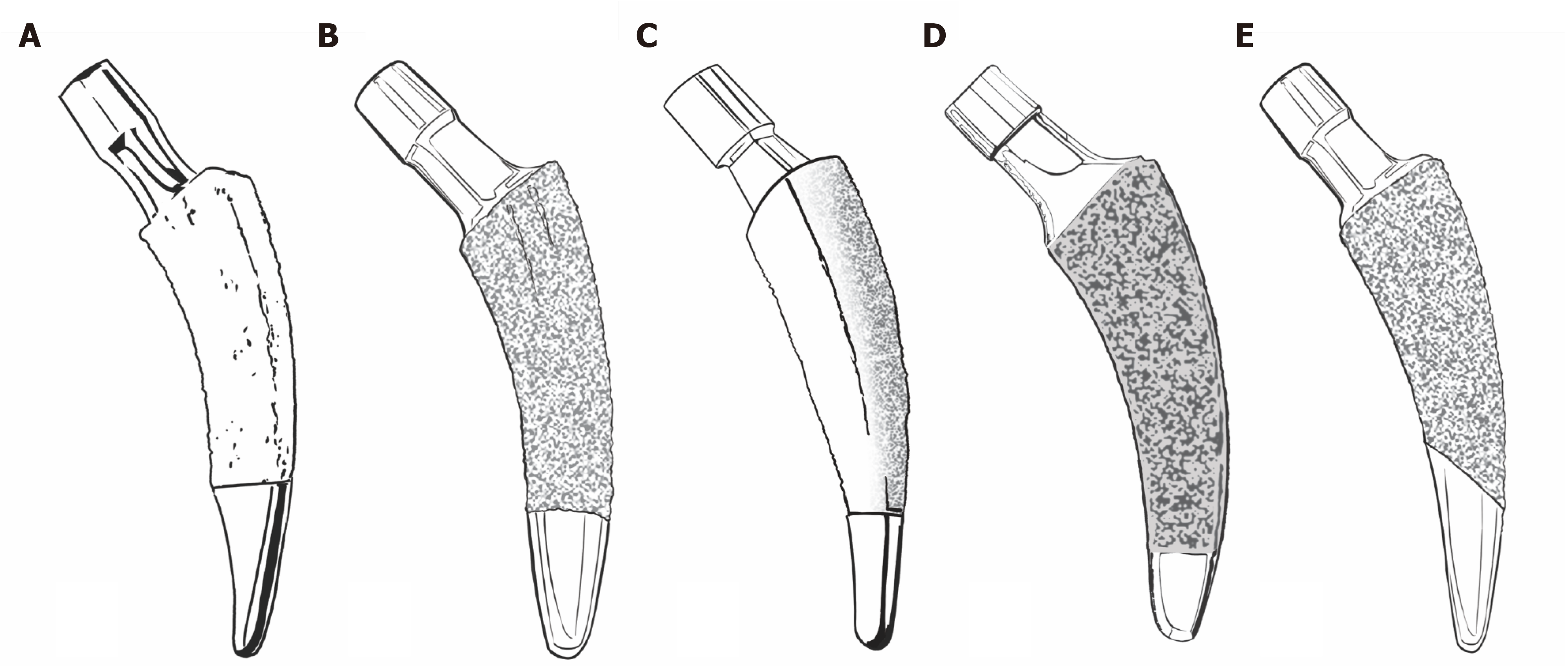
Figure 1 Common new-generation calcar-guided short stems.
A: Nanos stem (Smith&Nephew, Marl, Germany; on the market since 2004); B: MiniHip stem (Corin, Cirencester, UK; on the market since 2007); C: ColloMIS stem (Lima Corporate, Villanova di San Daniele del Friuli, Italy; on the market since 2009); D: optimys stem (Mathys Ltd., Bettlach, Switzerland; on the market since 2013); E: A2 stem (Artiqo, Luedinghausen, Germany; on the market since 2016).
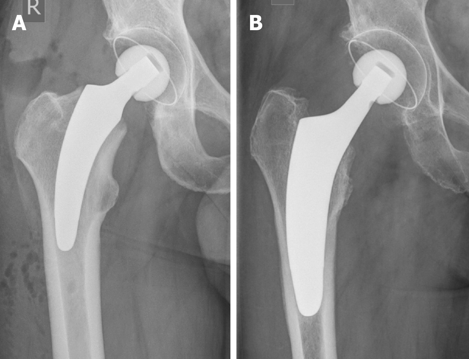
Figure 2 Introduction of a subclassification to account for the individualized positioning of group 2A and 2B stems, according to Khanuja etal[24].
A: Metaphyseal anchorage [for example group 2B(M)]; classical three-point anchoring; B: Meta-diaphyseal anchorage [group 2B(MD)]; additional fit-and-fill in the proximal diaphysis. Citation: Khanuja HS, Banerjee S, Jain D, Pivec R, Mont MA. Short bone-conserving stems in cementless hip arthroplasty. J Bone Joint Surg Am 2014; 96: 1742-1752.
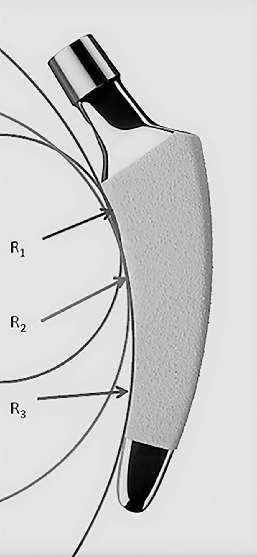
Figure 3 The design of modern calcar-guided short stems, with a rounded shape, is adapted to the medial anatomical calcar curve.
In the case of the optimys stem (Mathys Ltd., Bettlach, Switzerland), three different radii were used to design the curve (R1-3). (Copyright Mathys Ltd., Bettlach, Switzerland).
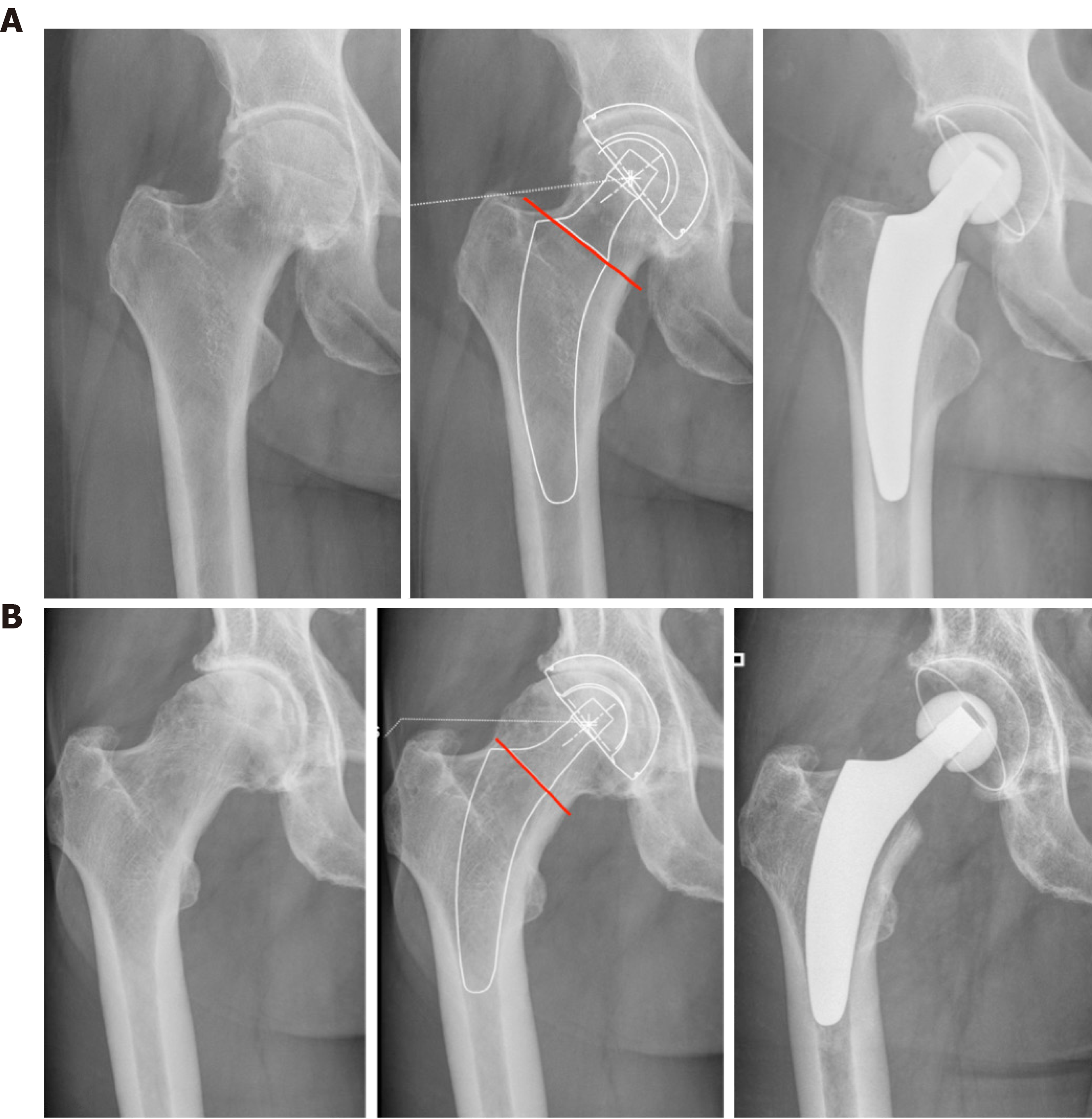
Figure 4 Individualized levels of resection, according to preoperative planning.
A: Valgus anatomy; B: Varus anatomy.
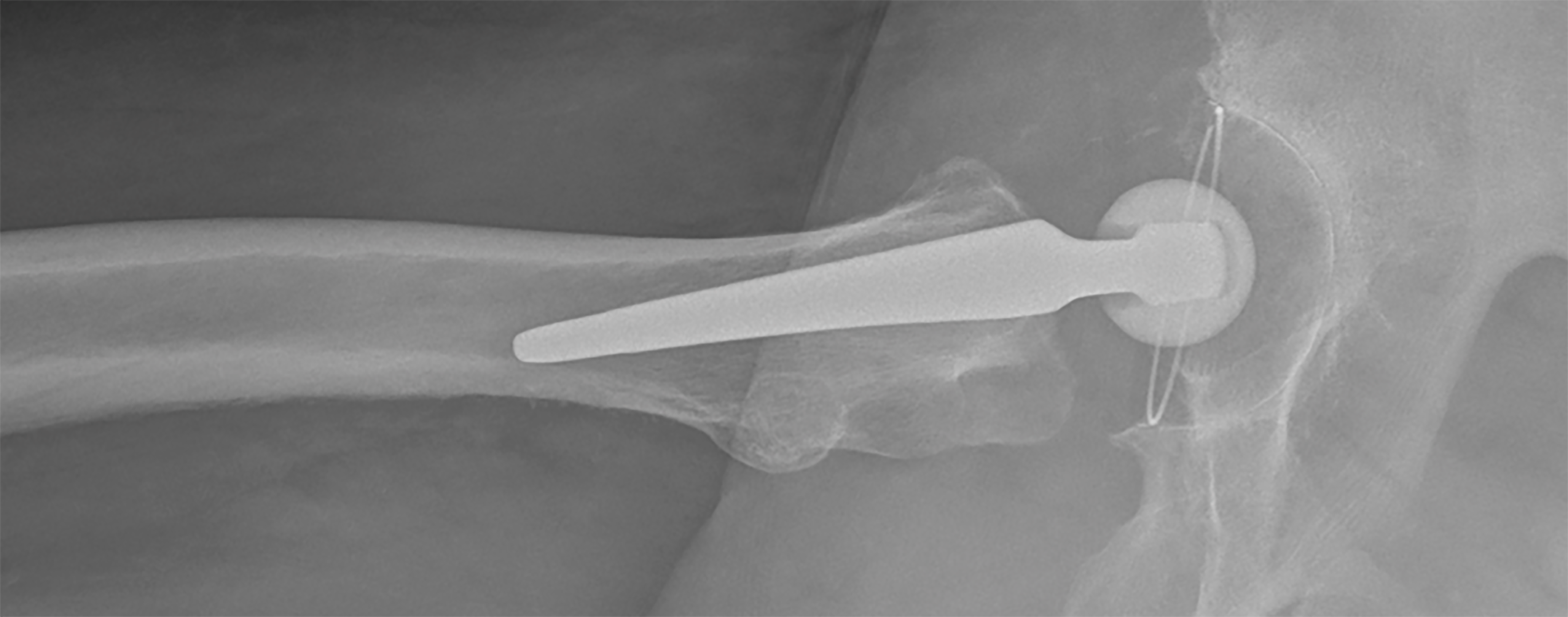
Figure 5 The natural anterior tilt of the femoral neck (in the second plane) can only be reconstructed using a short stem.
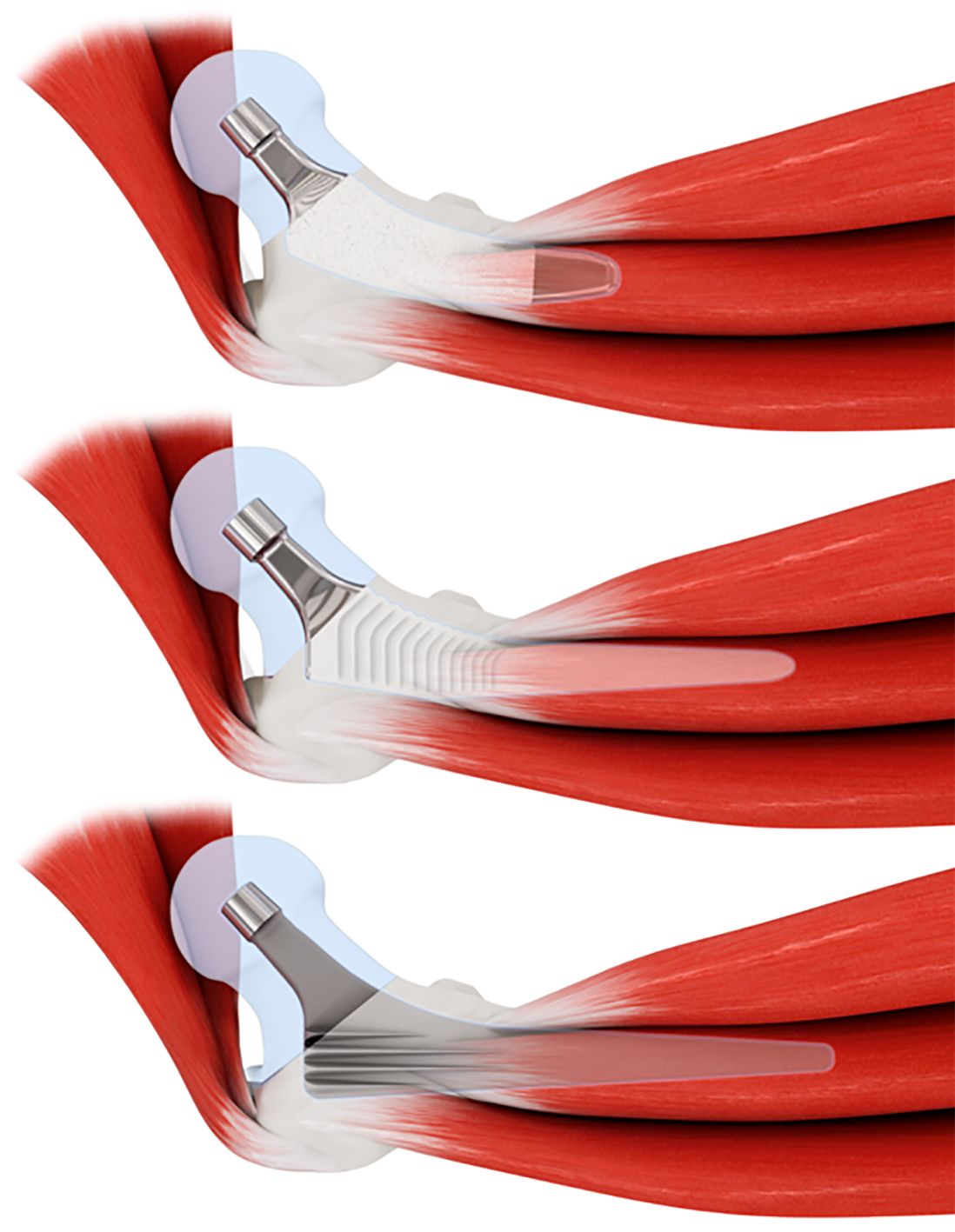
Figure 6 Using rounded short stems (top), minimally invasive approaches, without requiring transection or damage to the muscles, are facilitated compared with conventional stems (middle and bottom).
Copyright Mathys Ltd., Bettlach, Switzerland.
Figure 7 Metaphyseal anchoring using three-point fixation.
A: Mayo stem (Zimmer Biomet, Warsaw, Indiana, USA); B: Metha stem (Aesculap, Tuttlingen, Germany); C: Nanos stem (Smith&Nephew, Marl, Germany).
Figure 8 Characteristics of various short stem designs at different CCD angles, according to the classification of Groups A–E established by Kutzner etal[22,35].
A: Metha stem (Aesculap, Tuttlingen, Germany); additional diaphyseal anchorage is almost impossible to attain; B: Nanos stem (Smith&Nephew, Marl, Germany); narrowing in the distal part limits the option of additional diaphyseal anchorage; C: Optimys stem (Mathys Ltd., Bettlach, Switzerland); in neutral and valgus positions, an additional diaphyseal anchorage is possible, when intended. (Copyright Mathys Ltd., Bettlach, Switzerland).
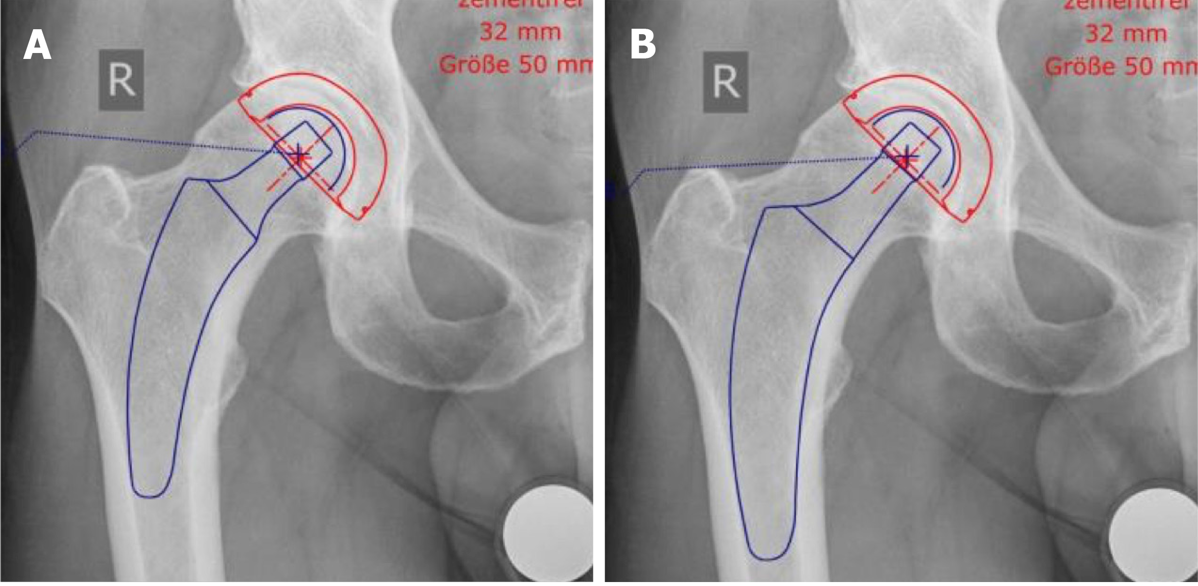
Figure 9 The same patient can be planned and treated by the application of metaphyseal anchorage or the addition of a fit-and-fill in the proximal diaphysis by valgization and upsizing, depending on the indication and bone quality.
A: Metaphyseal anchorage; B: Additional fit-and-fill in the proximal diaphysis.
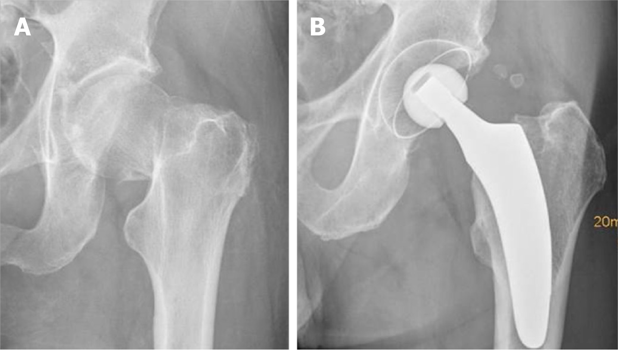
Figure 10 Calcar-guided short stem total hip arthroplasty in a case of osteonecrosis of the femoral head.
A: Preoperative; B: Postoperative.
Figure 11 Calcar-guided short stem total hip arthroplasty in a case of a femoral neck fracture.
A: Preoperative; B: Postoperative.
Figure 12 Prototype of a cemented calcar-guided short stem made out of polished steel (optimys stem, Mathys Ltd.
, Bettlach, Switzerland). Copyright Mathys Ltd., Bettlach, Switzerland.
- Citation: Kutzner KP. Calcar-guided short-stem total hip arthroplasty: Will it be the future standard? Review and perspectives. World J Orthop 2021; 12(8): 534-547
- URL: https://www.wjgnet.com/2218-5836/full/v12/i8/534.htm
- DOI: https://dx.doi.org/10.5312/wjo.v12.i8.534


