Copyright
©The Author(s) 2021.
World J Orthop. Jul 18, 2021; 12(7): 505-514
Published online Jul 18, 2021. doi: 10.5312/wjo.v12.i7.505
Published online Jul 18, 2021. doi: 10.5312/wjo.v12.i7.505
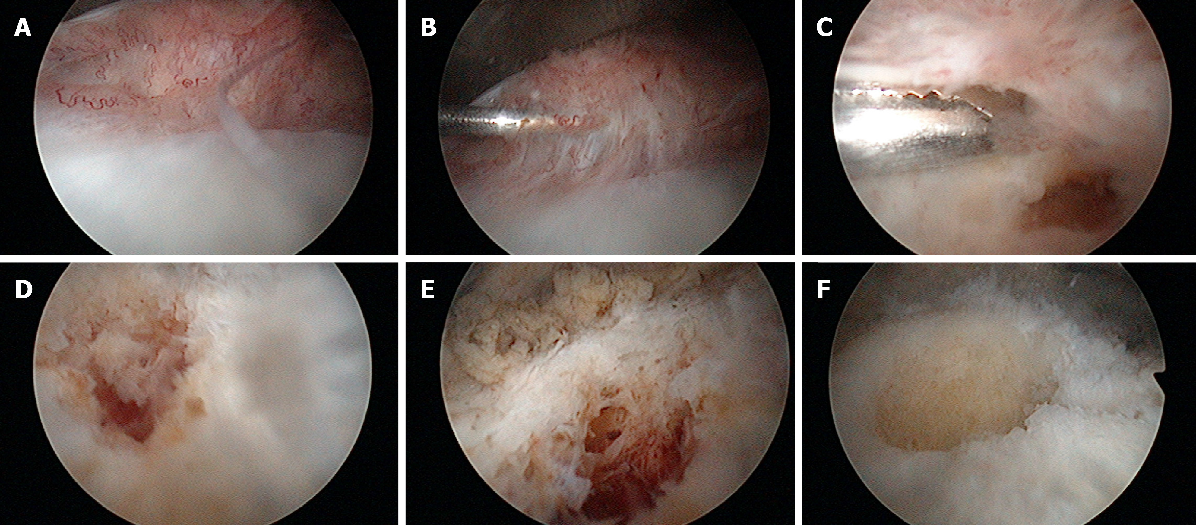
Figure 1 Sequential arthroscopic views of the anteromedial part of the femur above the cartilage with the arthroscope in the anterolateral portal.
A: synovial hyperplasia; B: Palpation of the elevated synovial tissue with a probe; C: Removal of the synovial tissue reveals the altered tissue; D and E: After partial removal, the extensiveness of the altered tissue is revealed; F: The remaining crater after complete removal of the lesion.
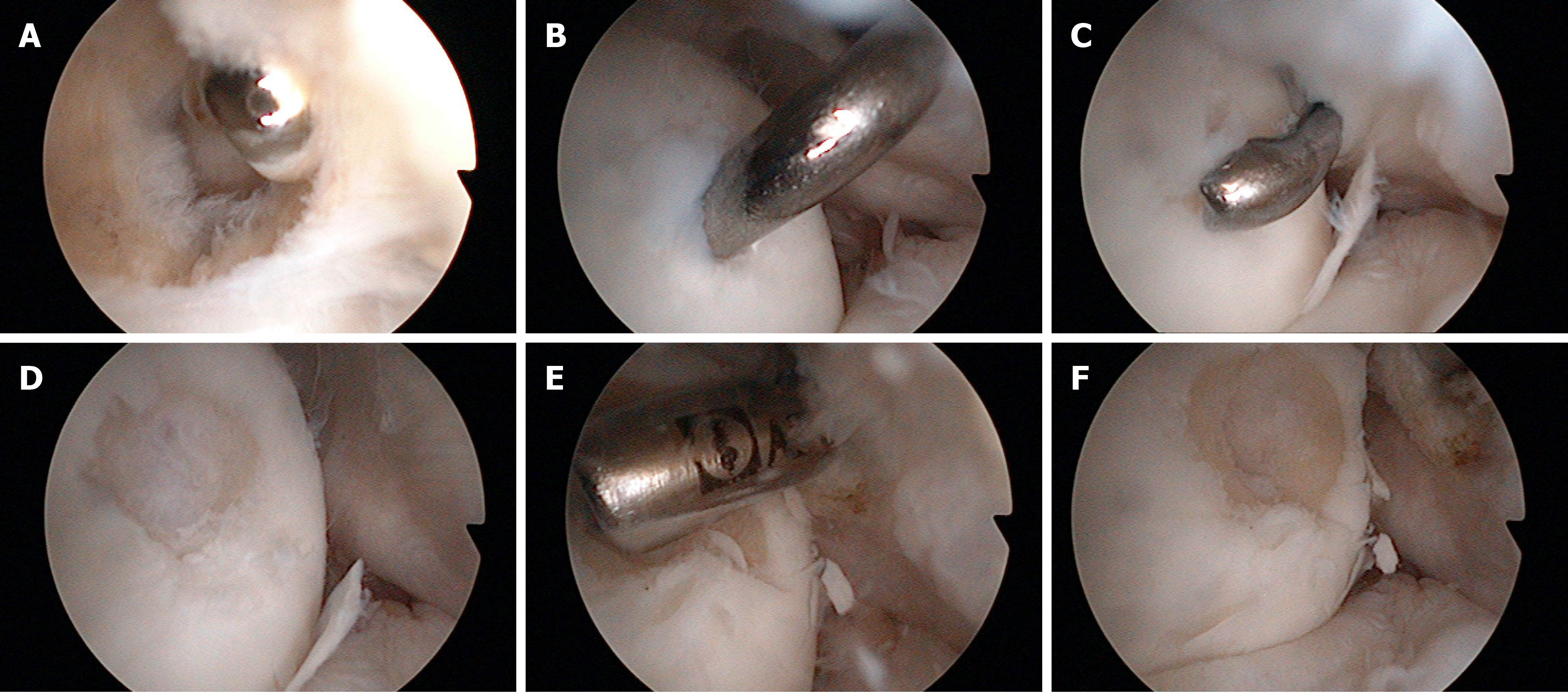
Figure 2 Sequential arthroscopic views of posterior part of lateral femoral condyle with the arthroscope positioned in the posteromedial portal.
A: Trans-septal approach; B: Palpation of the altered tissue with a probe from the posterolateral portal; C: Removal of the altered tissue with a cochlea; D: Visualization of the sclerotic bone after removal of altered tissue; E: Removal of the nidus and surrounding sclerosis with a motorized instrument; F: The remaining crater after complete removal of the lesion.
- Citation: Plečko M, Mahnik A, Dimnjaković D, Bojanić I. Arthroscopic removal as an effective treatment option for intra-articular osteoid osteoma of the knee. World J Orthop 2021; 12(7): 505-514
- URL: https://www.wjgnet.com/2218-5836/full/v12/i7/505.htm
- DOI: https://dx.doi.org/10.5312/wjo.v12.i7.505


