Copyright
©The Author(s) 2021.
World J Orthop. Jul 18, 2021; 12(7): 485-494
Published online Jul 18, 2021. doi: 10.5312/wjo.v12.i7.485
Published online Jul 18, 2021. doi: 10.5312/wjo.v12.i7.485
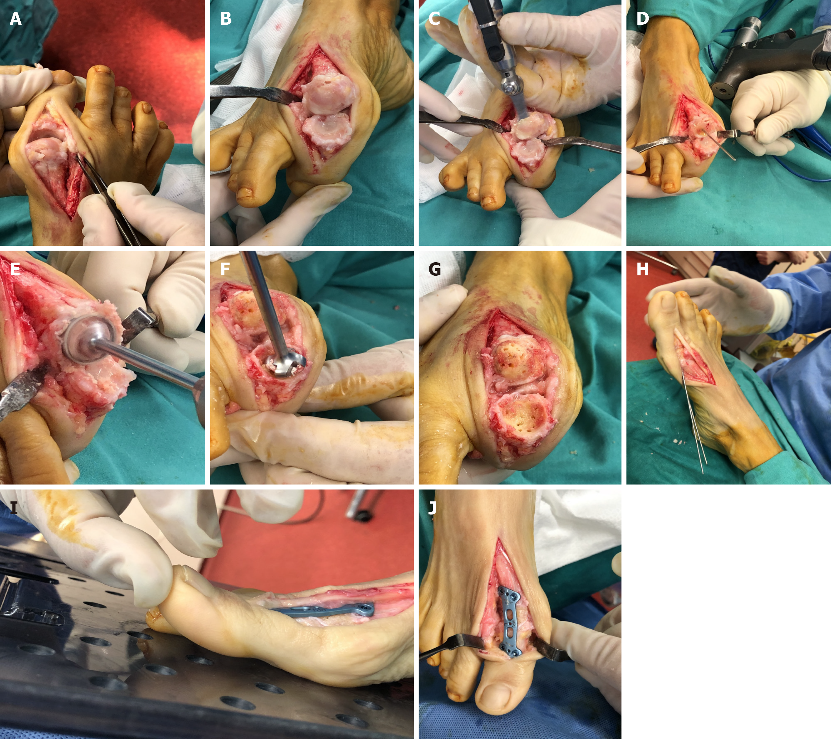
Figure 1 Surgical technique.
A: A dorsal midline approach provides adequate access to the joint; B: Extensor hallucis longus tendon is laterally retracted and adequate collateral ligament and capsule reflection provides excellent working space; C: A minimal flat cut is made to the head of the 1st metatarsal utilizing an oscillating saw; D: A guidewire is inserted to the center of the 1st metatarsal head; E and F: Hemispherical cup and cone reamers are used in order to remove the articular surfaces; G: Concave and convex surfaces of bleeding subchondral bone have been prepared with multiple perforations; H: Provisional fixation with two K- wires in the considered optimum position; I: The sagittal alignment is being checked intraoperatively using a flat tray simulating weight bearing. The 1st metatarsophalangeal joint is positioned in a way that allows the pulp of the great toe to rest 5-10 mm above the flat surface; J: Final position of the plate.
- Citation: Koutsouradis P, Savvidou OD, Stamatis ED. Arthrodesis of the first metatarsophalangeal joint: The “when and how”. World J Orthop 2021; 12(7): 485-494
- URL: https://www.wjgnet.com/2218-5836/full/v12/i7/485.htm
- DOI: https://dx.doi.org/10.5312/wjo.v12.i7.485


