Copyright
©The Author(s) 2021.
World J Orthop. May 18, 2021; 12(5): 329-337
Published online May 18, 2021. doi: 10.5312/wjo.v12.i5.329
Published online May 18, 2021. doi: 10.5312/wjo.v12.i5.329
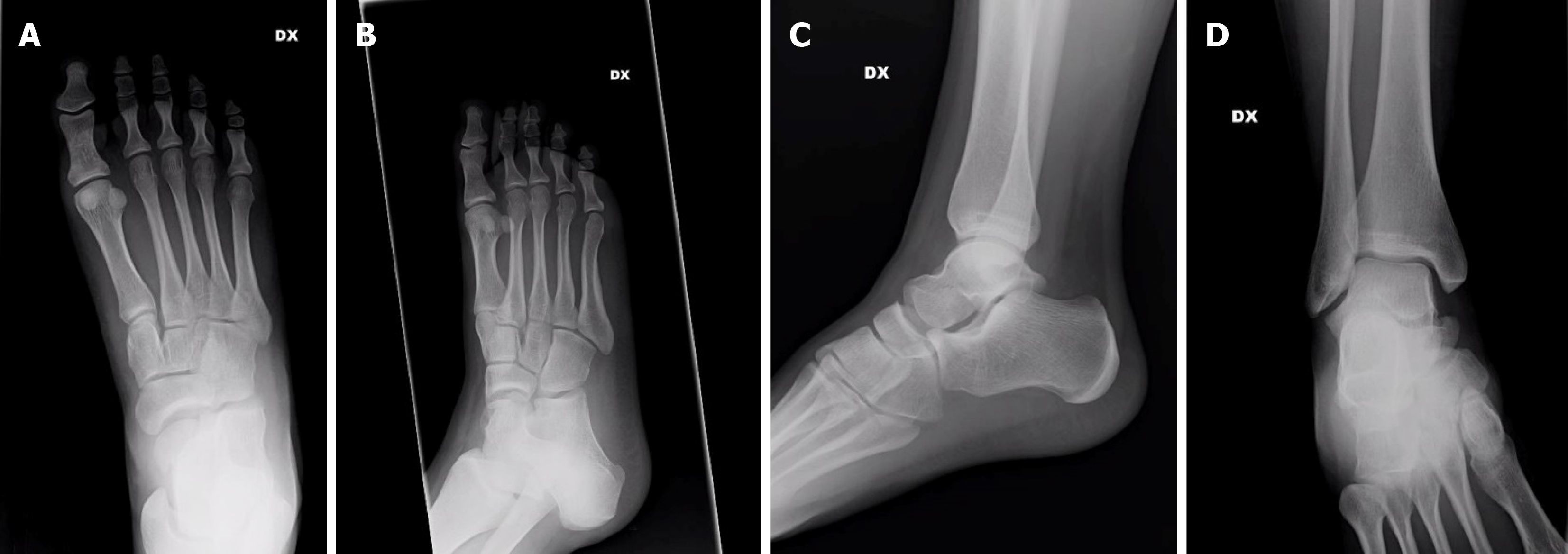
Figure 1 X-rays of the patient at the emergency department.
A: The foot was investigated by A-P view; B: Oblique view; C: Ankle on lateral view; and D: A-P view.
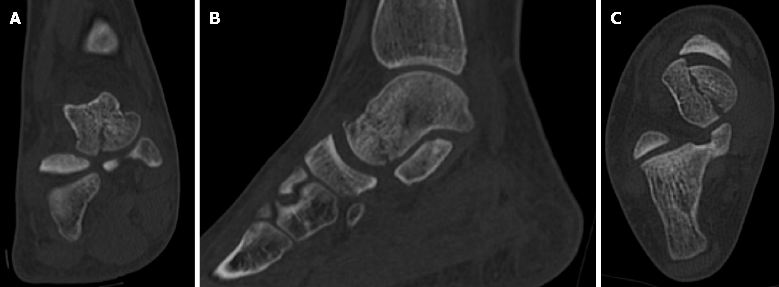
Figure 2 Pre-operative computed tomography images of the shear-type fracture.
A: Coronal, B: Sagittal; and C: Axial view are shown.
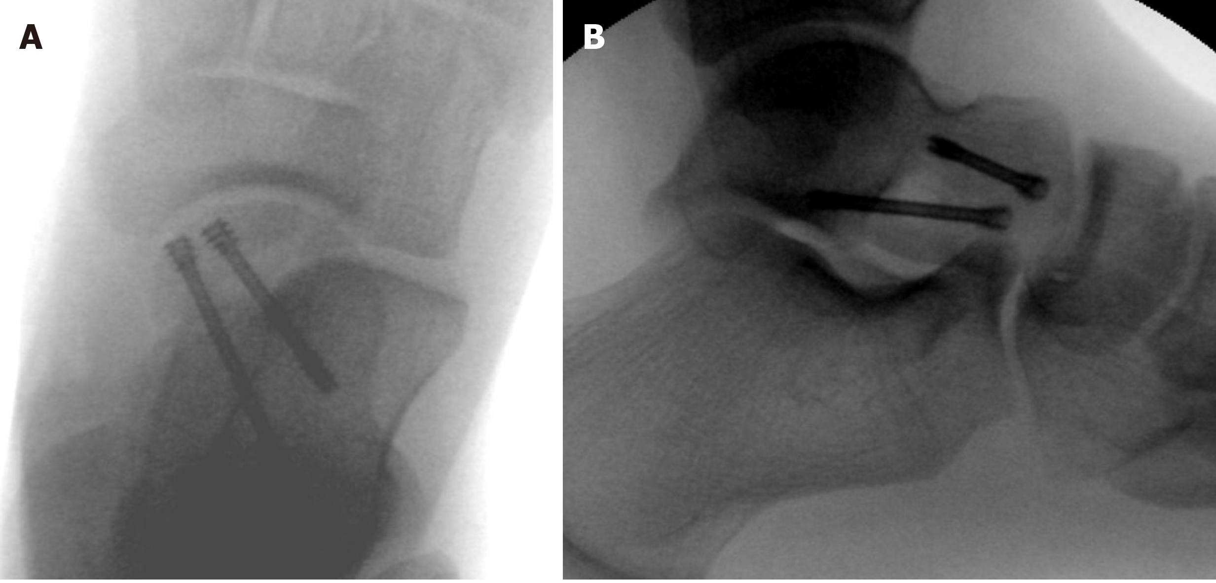
Figure 3 Intraoperative X-rays.
Two headless screws were used for fixation. A: Anteroposterior view; B: Lateral view.
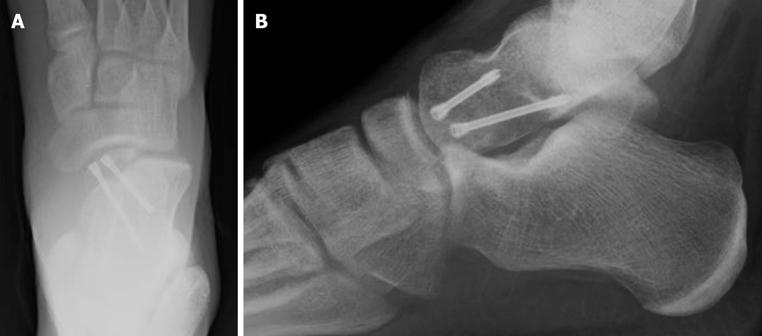
Figure 4 Plain films at one-month postoperative follow-up: the fracture is healing.
A: Anteroposterior view; B: Lateral view.
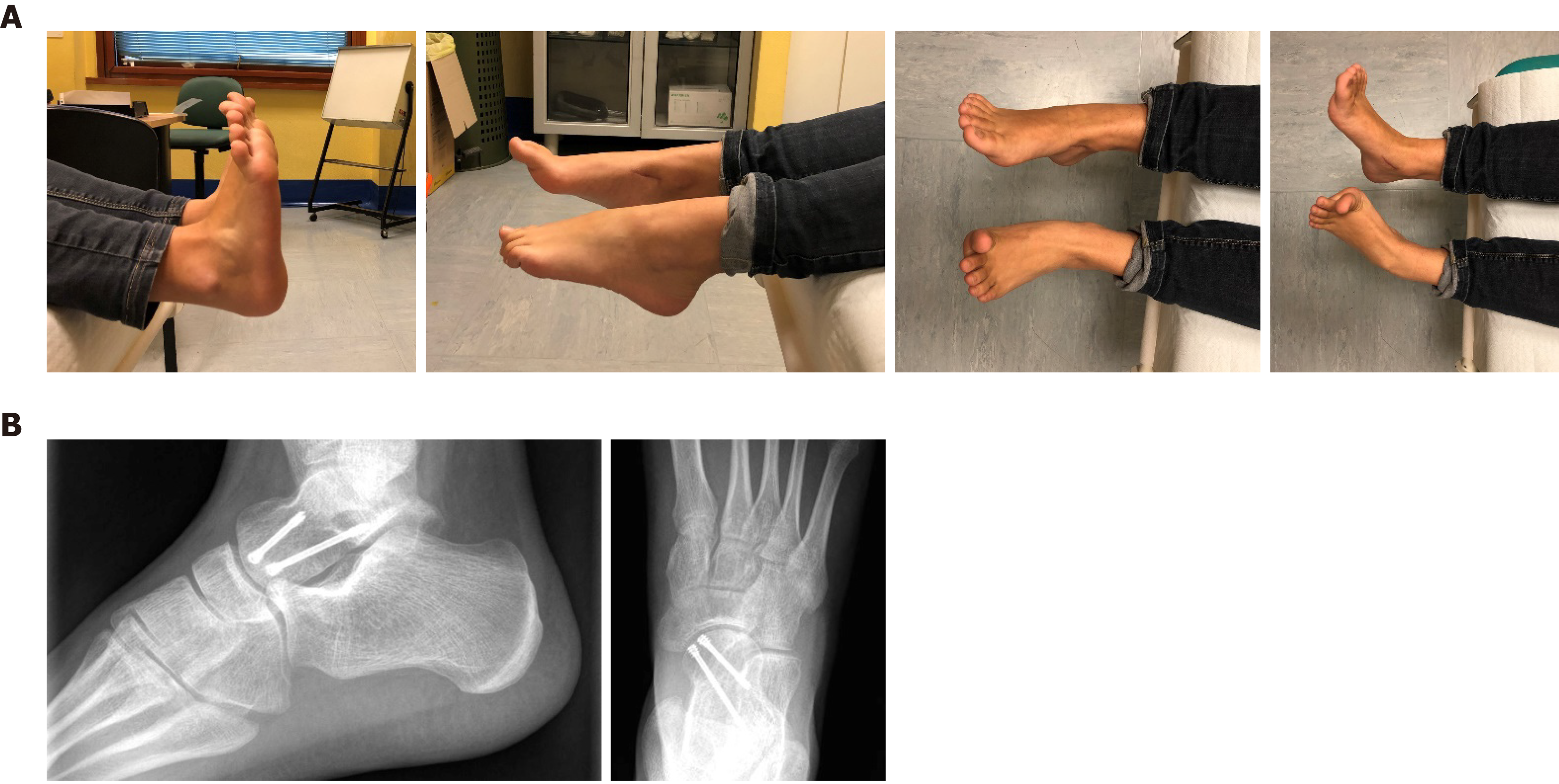
Figure 5 Follow-up at two months: the foot is shrunken and the surgical wound healed.
Plain films revealed a healed fracture. A: Clinical assessment; B: Lateral view; and C: Anteroposterior view.
Figure 6 Follow-up at six months: complete range of motion was restored.
A: Clinical assessment; and B: Fracture healed without osteonecrosis or osteoarthritis.
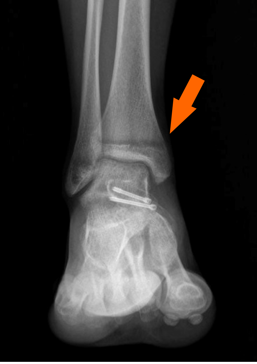
Figure 7 Postoperative plain film: The orange arrow shows that the physis of the distal tibia has started to close.
- Citation: Monestier L, Riva G, Faoro L, Surace MF. Rare shear-type fracture of the talar head in a thirteen-year-old child — Is this a transitional fracture: A case report and review of the literature. World J Orthop 2021; 12(5): 329-337
- URL: https://www.wjgnet.com/2218-5836/full/v12/i5/329.htm
- DOI: https://dx.doi.org/10.5312/wjo.v12.i5.329


