Copyright
©The Author(s) 2021.
World J Orthop. Mar 18, 2021; 12(3): 129-139
Published online Mar 18, 2021. doi: 10.5312/wjo.v12.i3.129
Published online Mar 18, 2021. doi: 10.5312/wjo.v12.i3.129
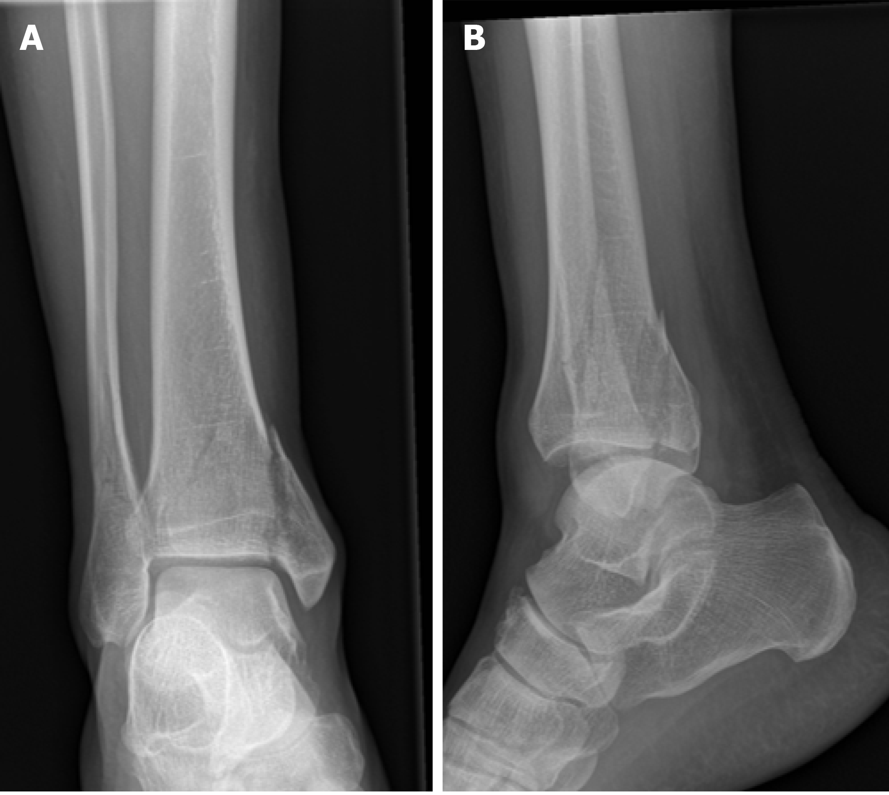
Figure 1 Transsyndesmotic fracture (44-B3).
A: Anteroposterior; B: Lateral view.
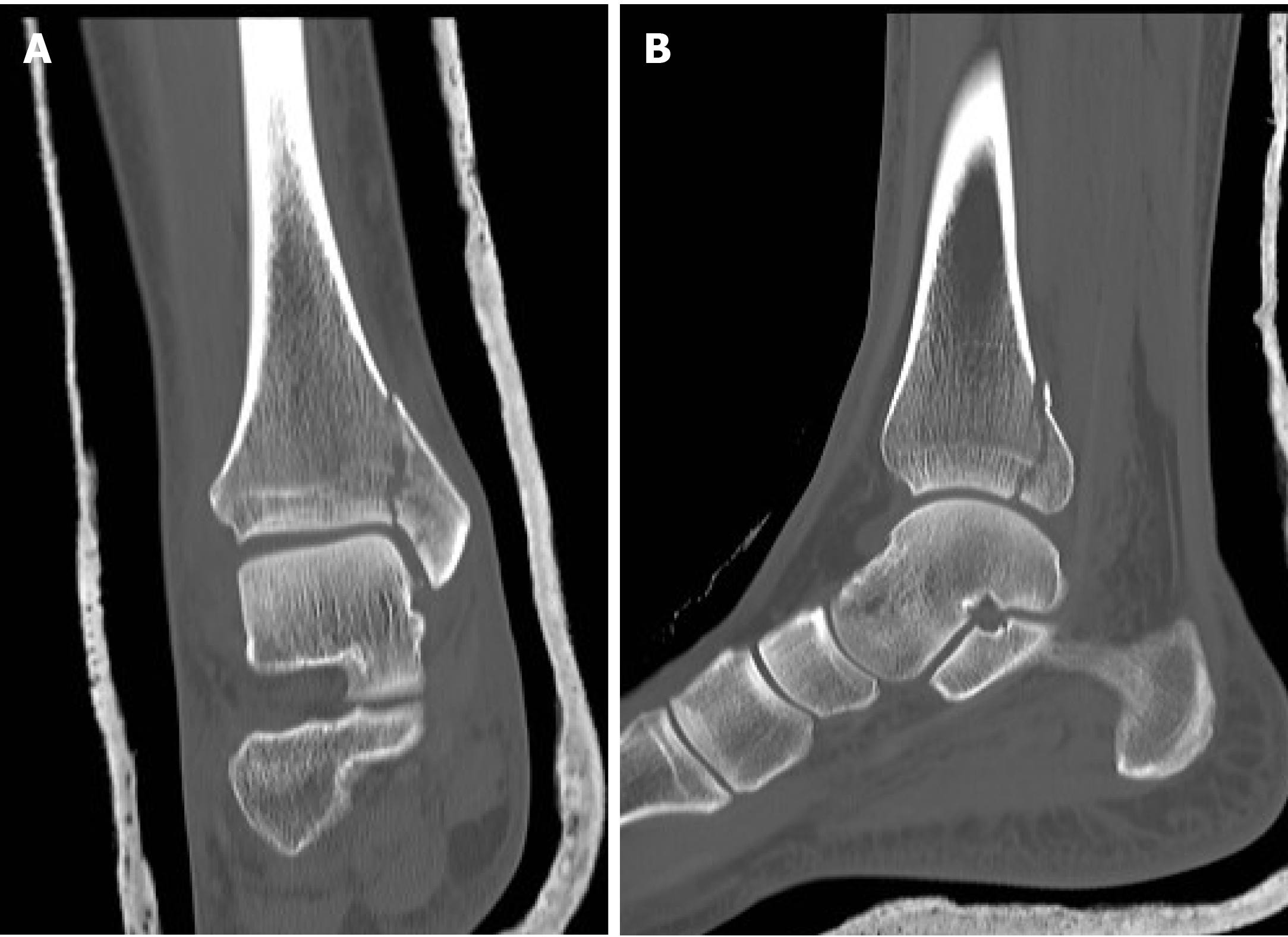
Figure 2 Computed tomography scans of the coronal plane and sagittal plane allow detection for the best screws direction.
A: Coronal plane; B: Sagittal plane.
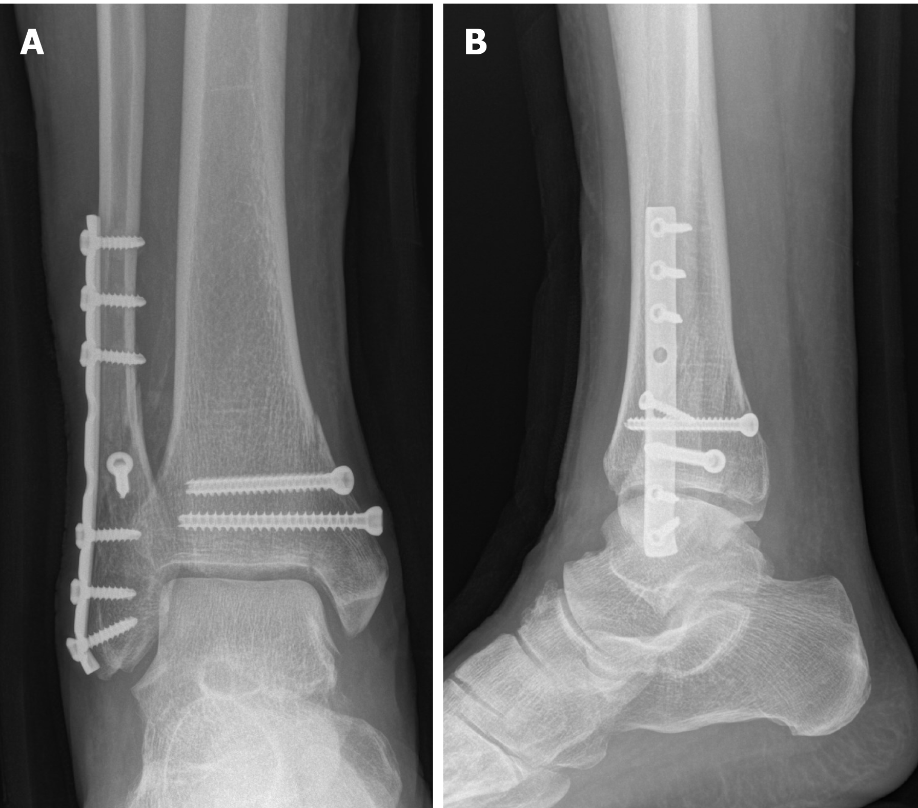
Figure 3 Postoperative X-rays in the anteroposterior and lateral view.
Fractures treated with plate and screw fixation. A: Anteroposterior view; B: Lateral view.
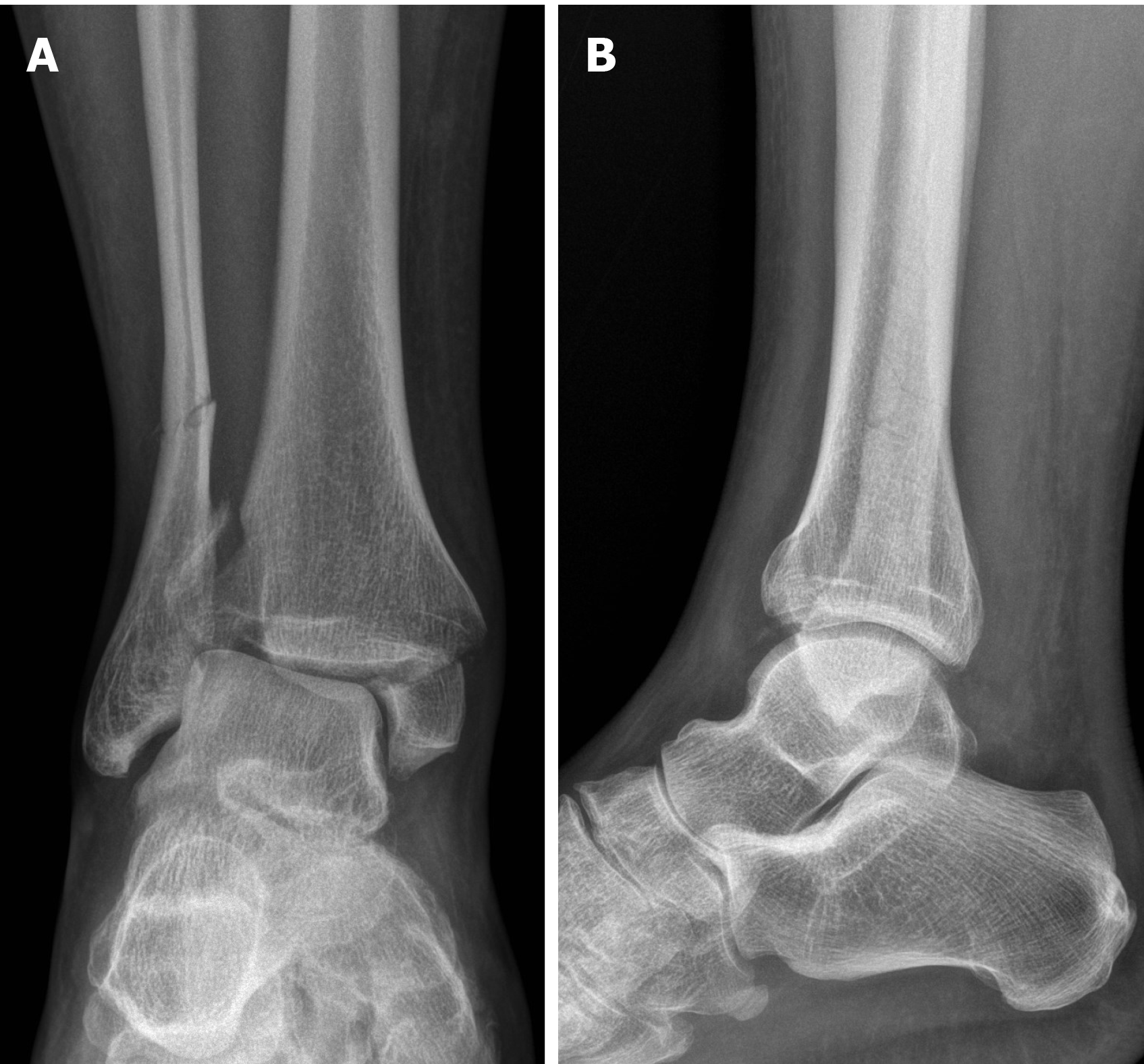
Figure 4 Suprasyndesmotic fracture (44-C2).
A: Anteroposterior; B: Lateral view.
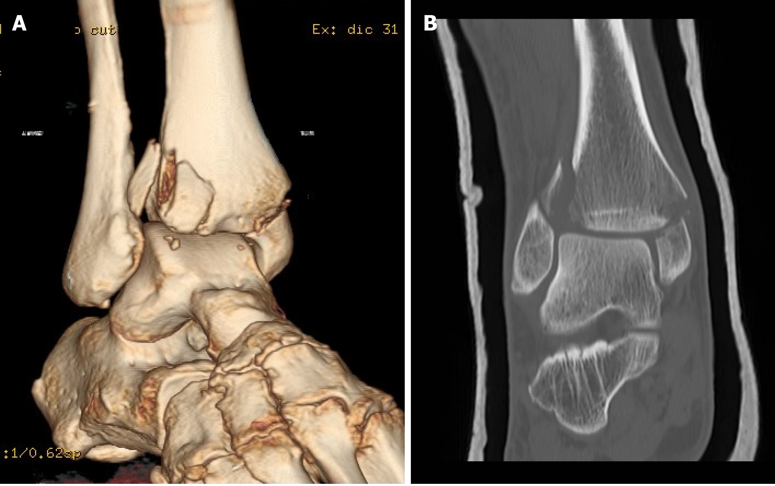
Figure 5 Computed tomography scans shows the involvement of the Tillaux-Chaput fragment.
A and B: Tillaux-Chaput fragment.
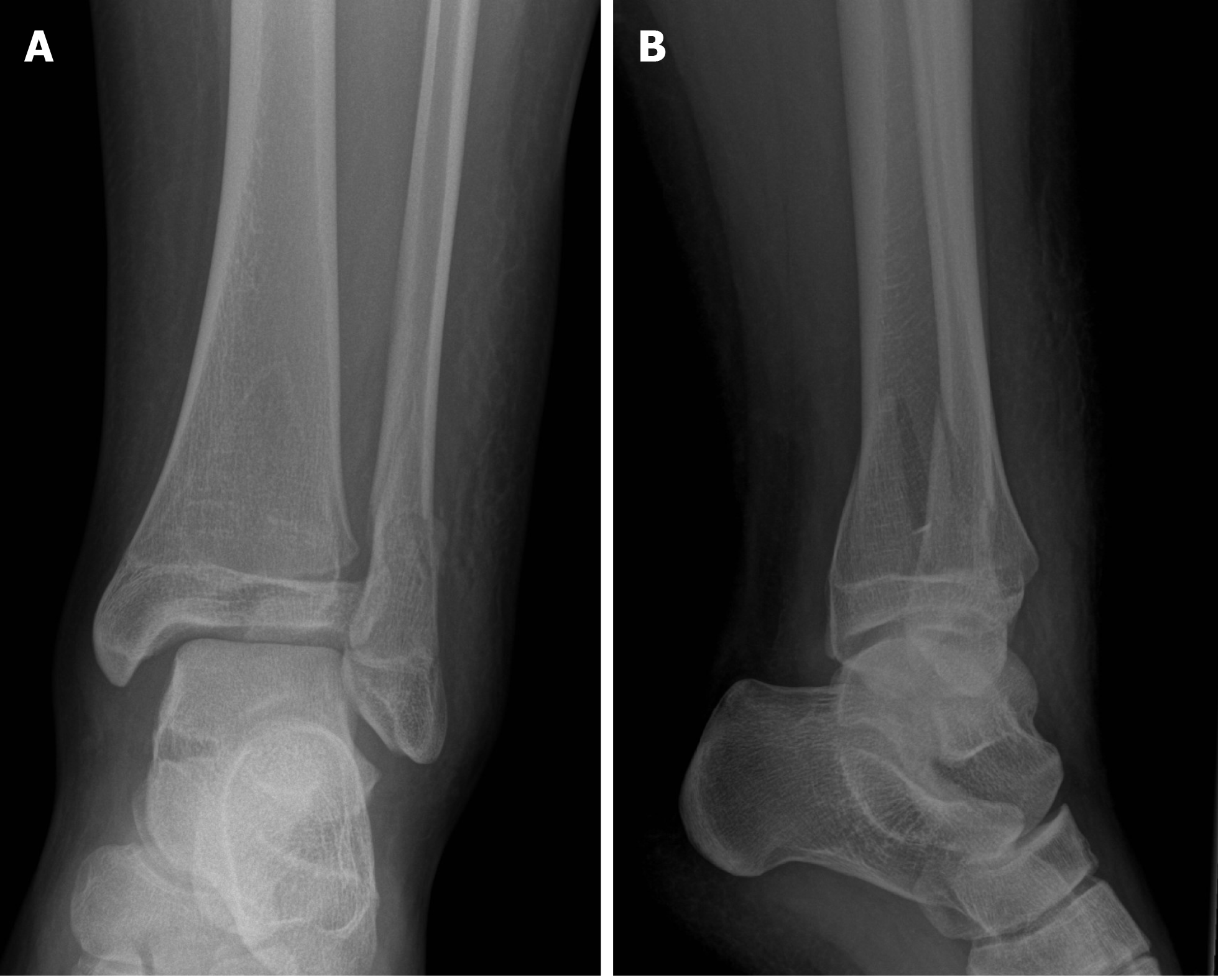
Figure 6 Ankle fracture in adolescent.
A: Anteroposterior view; B: Lateral view.
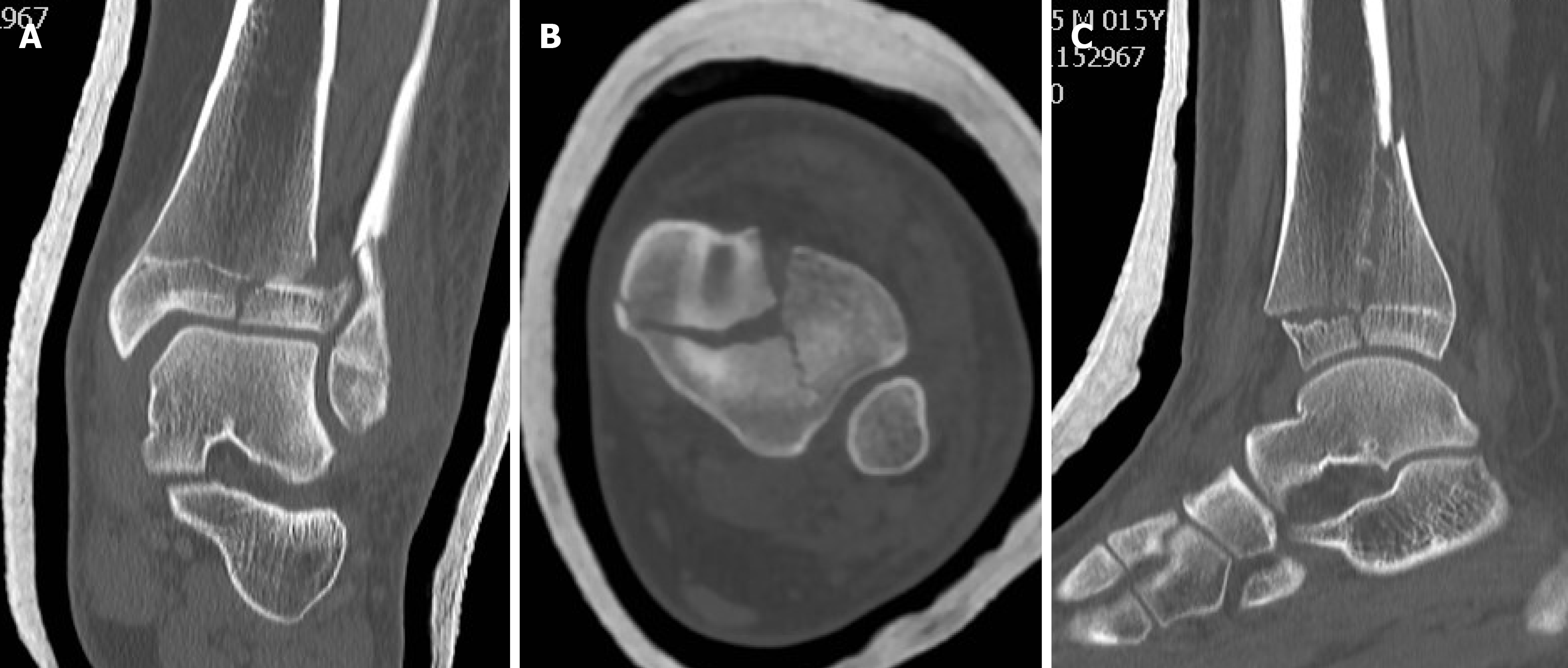
Figure 7 Computed tomography scan shows a triplane fracture.
A: Coronal plane; B: Axial plane; C: Sagittal plane.
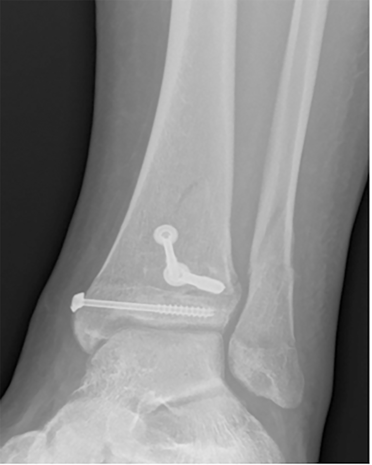
Figure 8 Postoperative X-rays in the anteroposterior view.
Fractures treated with screws.
- Citation: Tarallo L, Micheloni GM, Mazzi M, Rebeccato A, Novi M, Catani F. Advantages of preoperative planning using computed tomography scan for treatment of malleolar ankle fractures. World J Orthop 2021; 12(3): 129-139
- URL: https://www.wjgnet.com/2218-5836/full/v12/i3/129.htm
- DOI: https://dx.doi.org/10.5312/wjo.v12.i3.129


