Copyright
©The Author(s) 2021.
World J Orthop. Dec 18, 2021; 12(12): 970-982
Published online Dec 18, 2021. doi: 10.5312/wjo.v12.i12.970
Published online Dec 18, 2021. doi: 10.5312/wjo.v12.i12.970
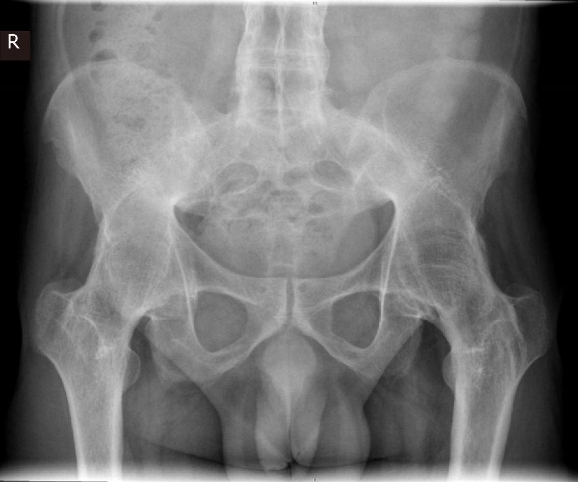
Figure 1 Bilateral fused hips with ankylosing spondylitis in a 43-year-old male at total hip arthroplasty-pre op.
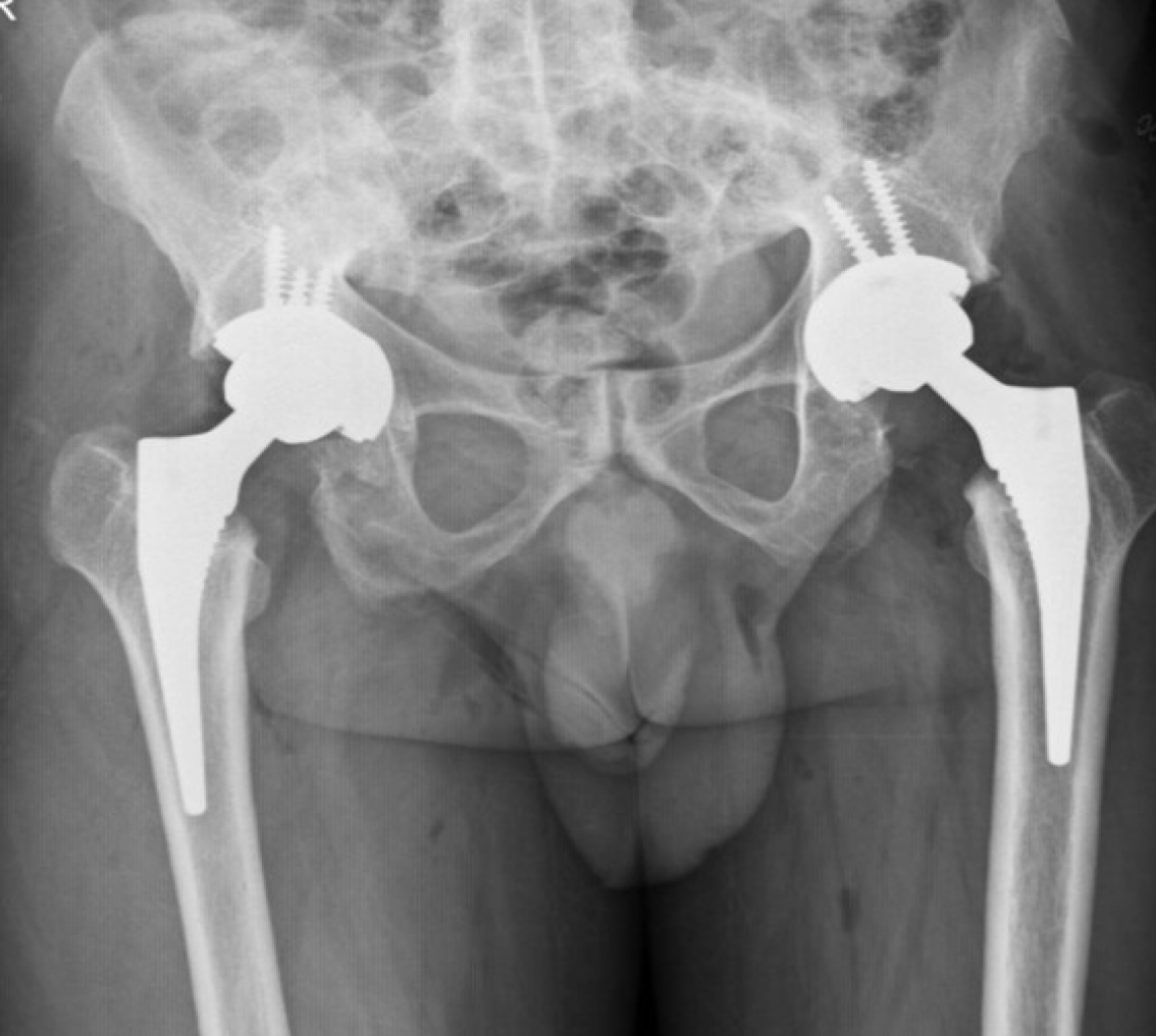
Figure 2 Bilateral fused hips-post op bilateral total hip arthroplasty (Pre op Figure 3) with cementless fixation in 43-year-old male.
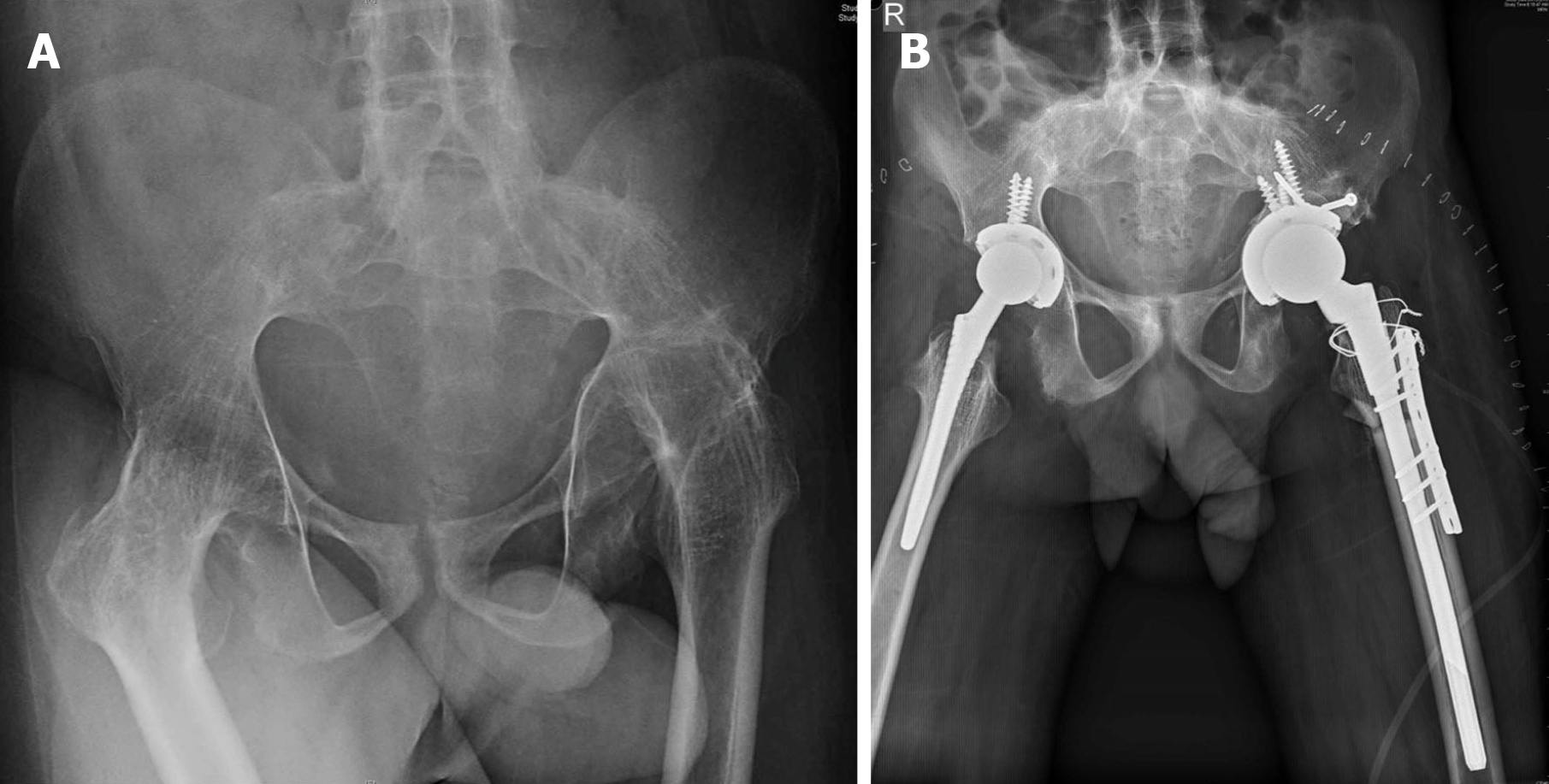
Figure 3 Bilateral fused hips and ankylosing spondylitis in a 31-year-old male.
A: Proximal migration of the left hip with femoral head and acetabular type 3b acetabular deficiency; B: Bilateral total hip arthroplasty with left hip femoral shortening. Acetabulum medial wall fracture, defect managed with bone graft and screws for superior augmentation. Sacral slope wire for proximal femur incomplete split, femur plate for additional rotational stability.
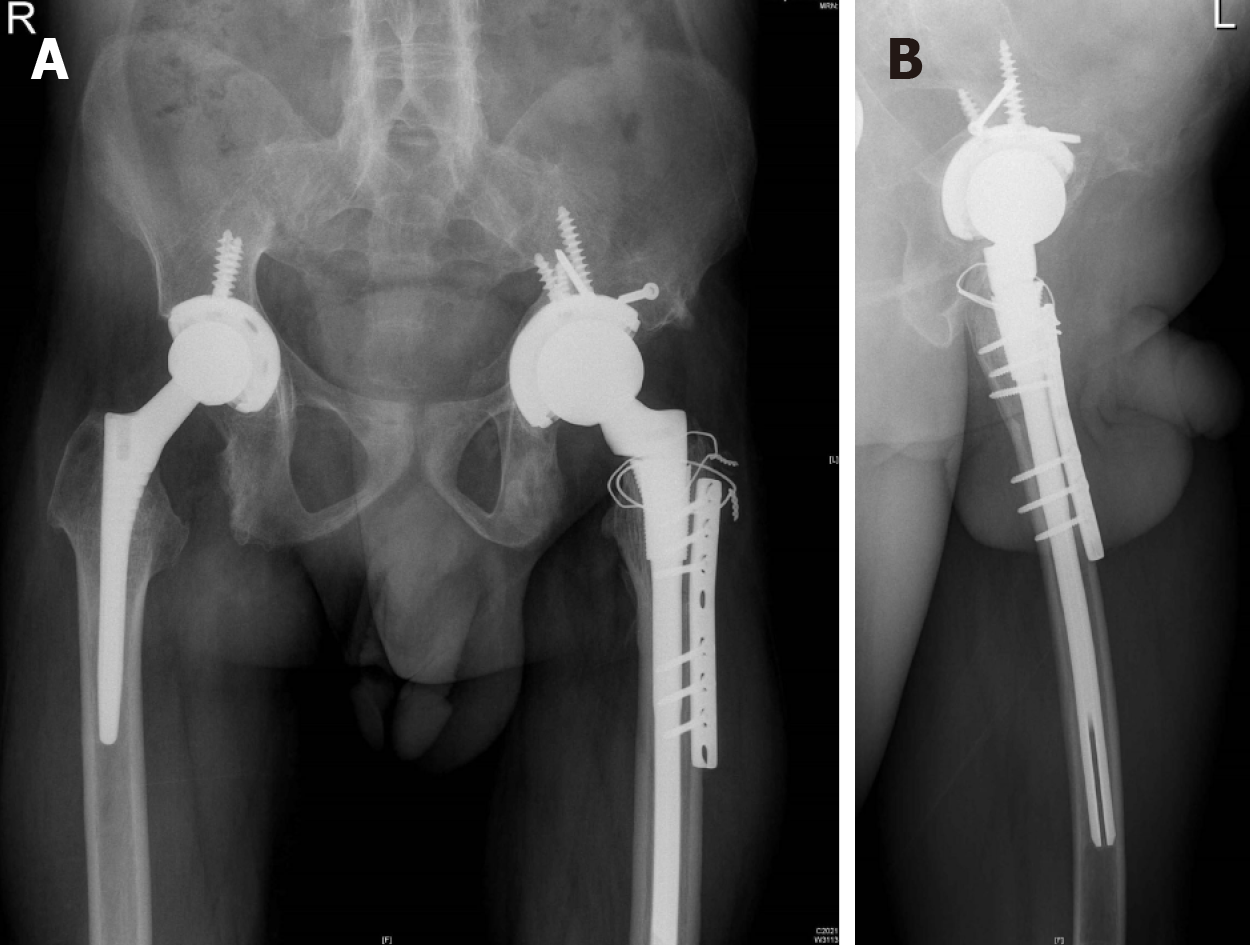
Figure 4 Follow up bilateral fused hips total hip arthroplasty in 31-year-old male with proximal migration left hip (pre op Figure 3).
A: 15 mo follow-up with osteotomy site union, acetabulum graft well united; B: Lateral view confirming osteotomy site union.
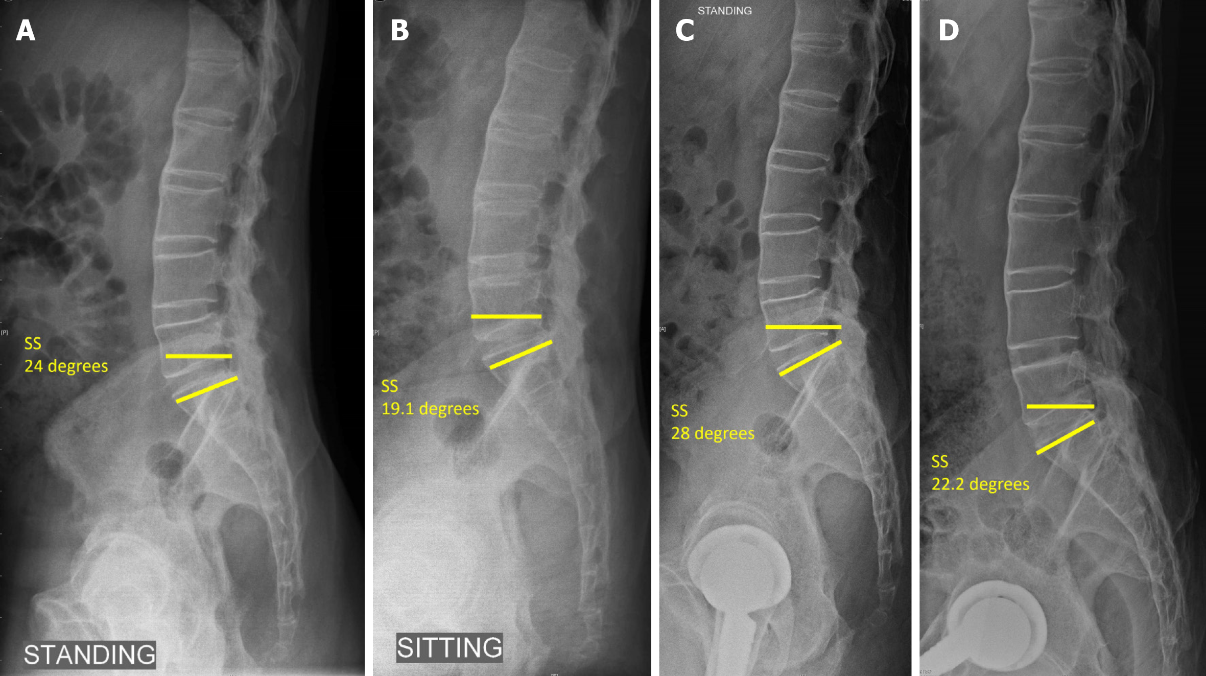
Figure 5 Lateral lumbosacral spine radiographs.
A: Pre op standing; B: Sitting compared to; C: Post op standing; D: Sitting showing the change in sacral slope < 10 degrees with reduced sacral slope indicating posterior pelvic tilt and stuck sitting pattern in ankylosing spondylitis (sacral slope < 30 degrees on sitting and standing typical of stuck sitting pattern). SS: Sacral slope.
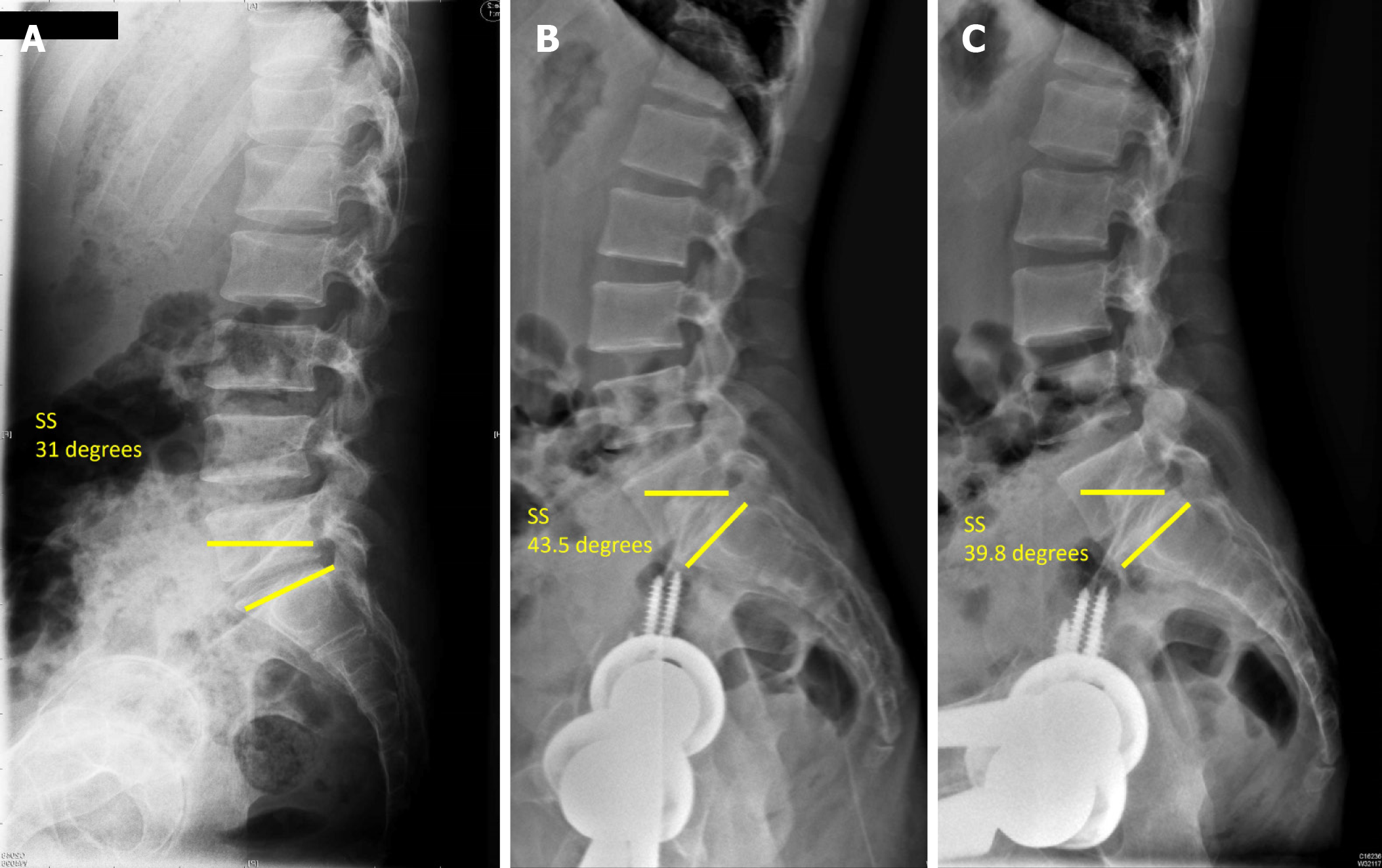
Figure 6 Spinopelvic mobility in a 51-year-old male with flexion deformity and inability to sit comfortably prior to total hip arthroplasty, pre op and 29 mo post bilateral total hip arthroplasty ankylosing spondylitis.
A: Pre op sacral slope (SS) standing; B: Post total hip arthroplasty SS standing; C: Sitting SS > 30 degrees sitting and standing demonstrates the stuck standing pattern. SS: Sacral slope.
Figure 7 Bilateral hip protrusion in 48-year-old male with ankylosing spondylitis and fused sacroiliac joints.
A: Pre op bilateral stiff hips; B: Post op total hip arthroplasty with bone grafting (autograft) reverse reaming for graft impaction; C: 1-year follow-up with graft integration.
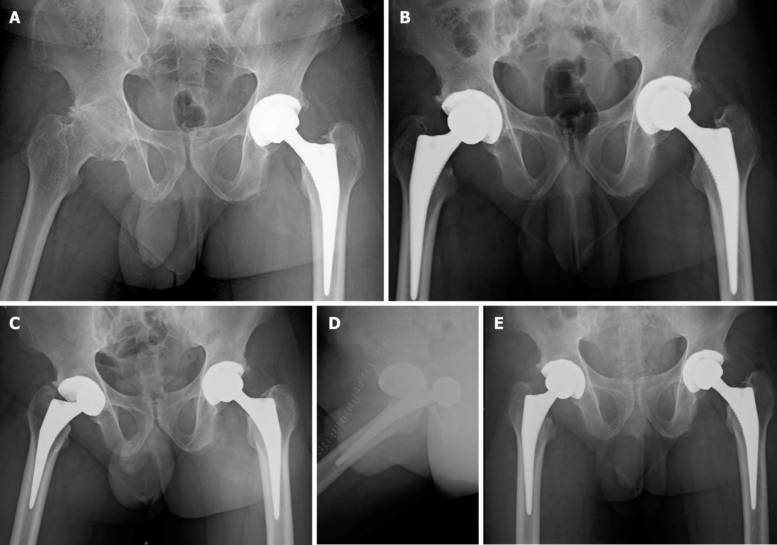
Figure 8 Left total hip arthroplasty with right hip arthritis in a 34-year-old male with ankylosing spondylitis.
A: Pre op stiff right hip; B: Post op right total hip arthroplasty; C: With dislocation at day 5 following a fall; D: Lateral view; E: 1-year follow-up after closed reduction.
Figure 9 Bilateral hip ankylosis in a 33-year-old male with ankylosing spondylitis.
A: Pre op total hip arthroplasty, 12-year post op fracture fixation left proximal femur; B: Post op bilateral total hip arthroplasty; C: Follow-up with Brooker grade 3 heterotopic ossification left hip and good hip function (Harris hip score improved from 34 to 81 at 24 mo follow-up).
- Citation: Oommen AT, Hariharan TD, Chandy VJ, Poonnoose PM, A AS, Kuruvilla RS, Timothy J. Total hip arthroplasty in fused hips with spine stiffness in ankylosing spondylitis. World J Orthop 2021; 12(12): 970-982
- URL: https://www.wjgnet.com/2218-5836/full/v12/i12/970.htm
- DOI: https://dx.doi.org/10.5312/wjo.v12.i12.970


