Copyright
©The Author(s) 2020.
World J Orthop. Mar 18, 2020; 11(3): 145-157
Published online Mar 18, 2020. doi: 10.5312/wjo.v11.i3.145
Published online Mar 18, 2020. doi: 10.5312/wjo.v11.i3.145
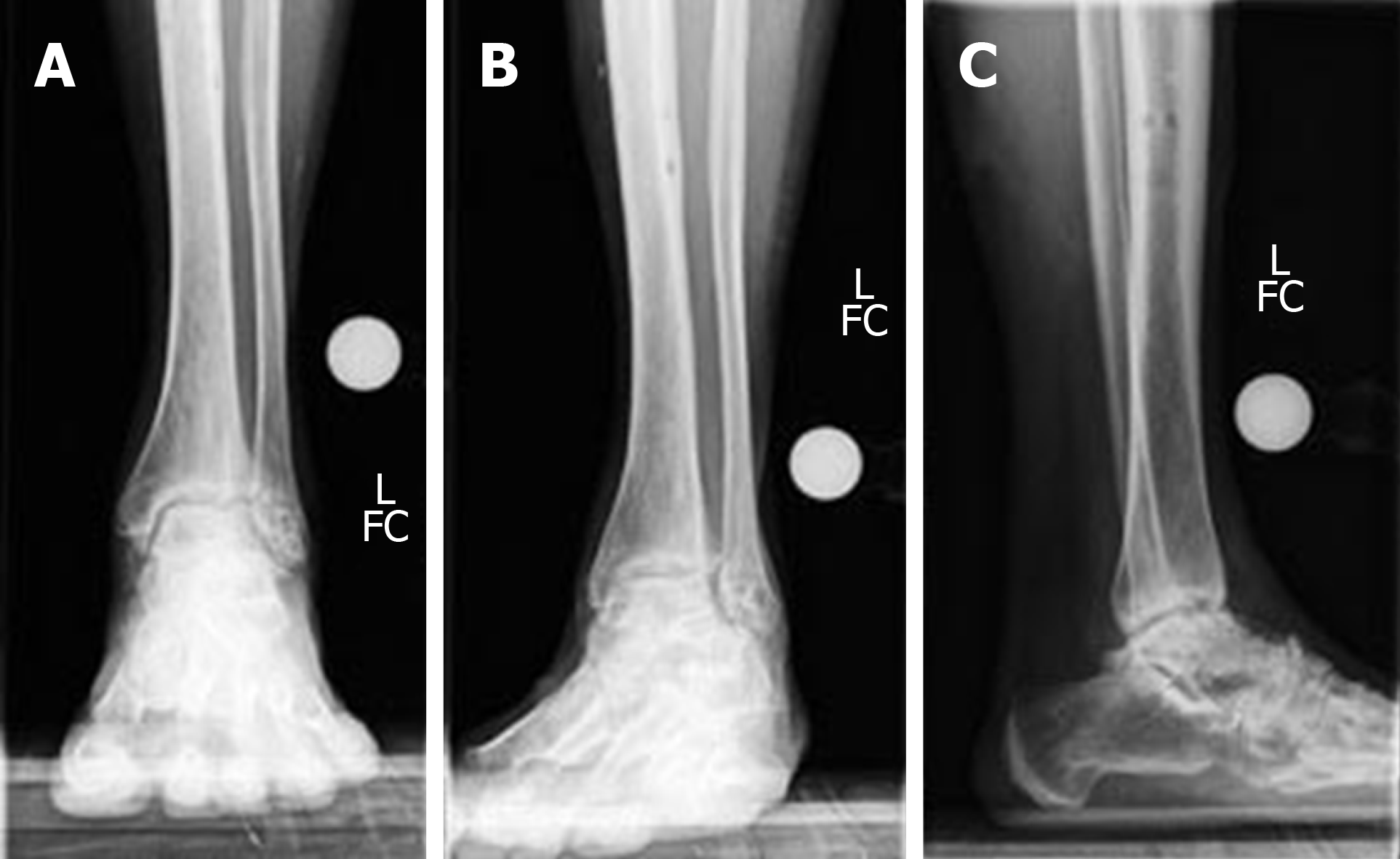
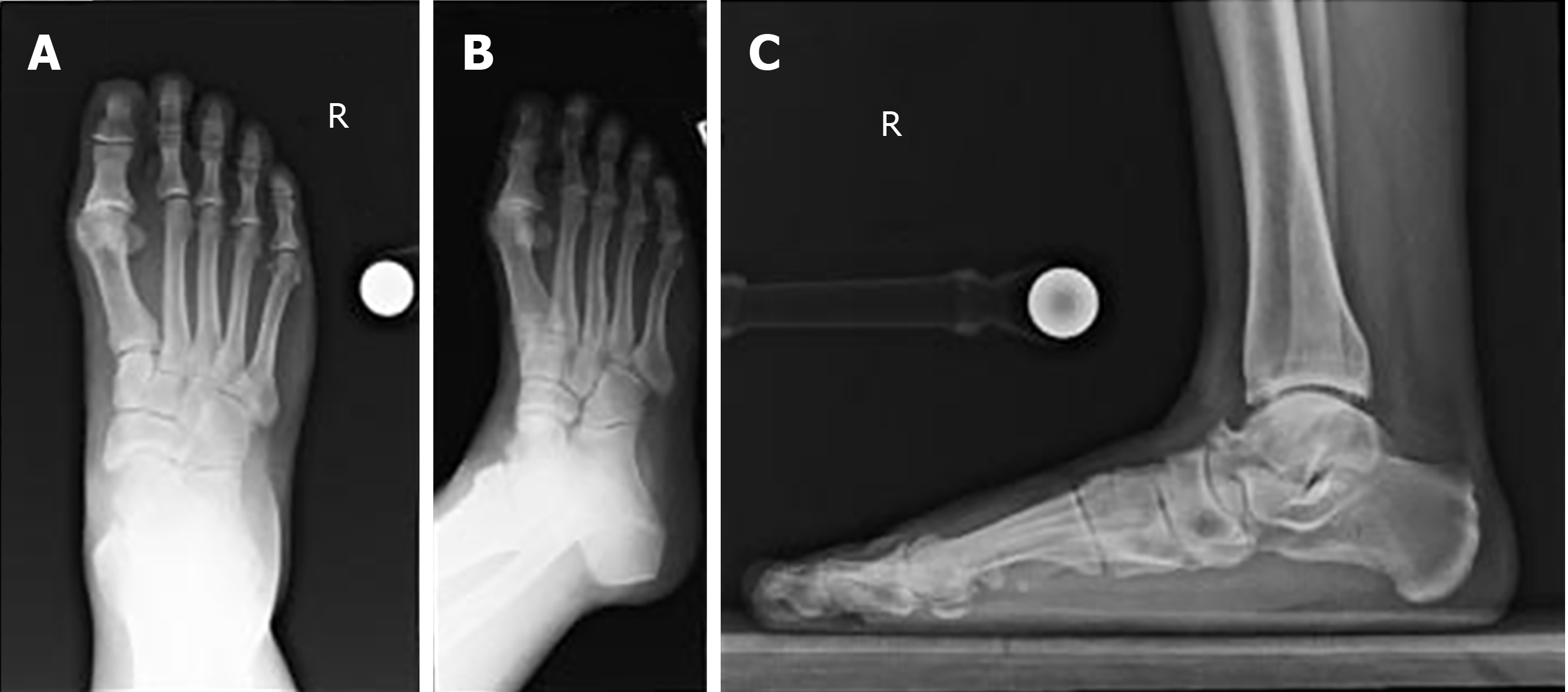
Figure 1 Pre-operative standing X-rays demonstrating post-traumatic osteoarthritis of the ankle joint.
A: Anterior-Posterior view; B: Mortise view; C: Lateral view.
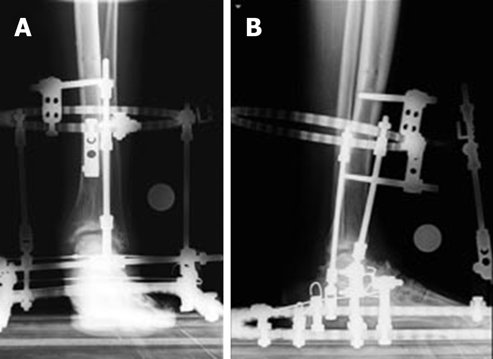
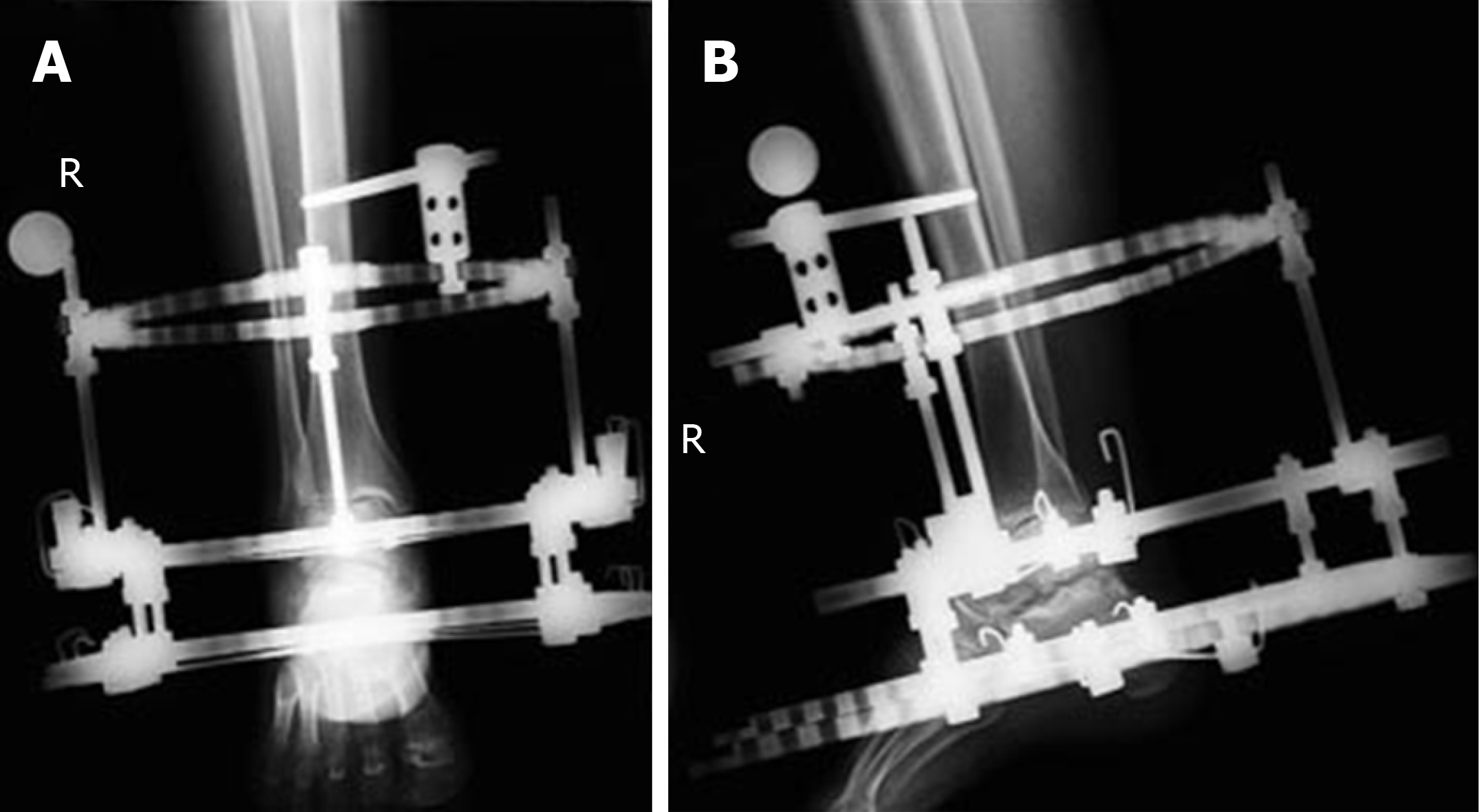
Figure 2 Post-operative standing X-rays demonstrating distracted ankle joint and distraction frame.
A: Anterior-Posterior view; B: Lateral view.
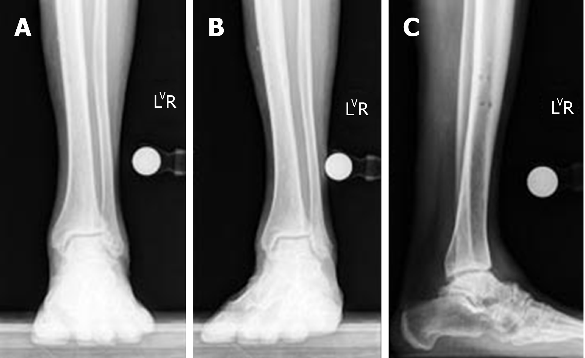
Figure 3 One-year post-operative standing X-rays demonstrating improved ankle joint space and articular surface after distraction.
A: Anterior-Posterior view; B: Mortise view; C: Lateral view.
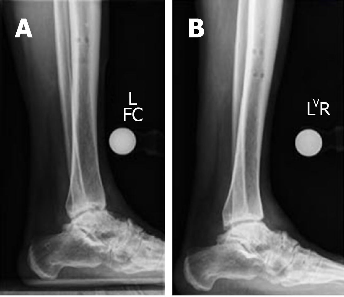
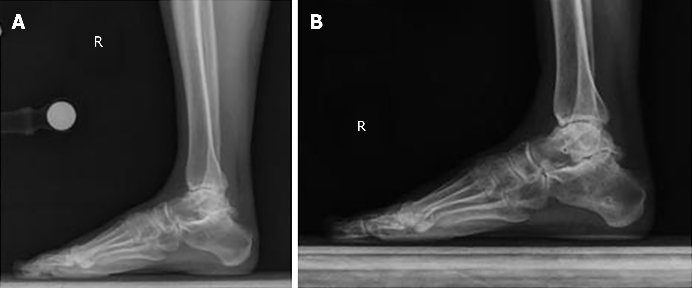
Figure 4 Comparison of pre and one-year post-operative standing X-rays demonstrating improved ankle joint space and articular surface after distraction.
A: Lateral view, pre-operative; B: Lateral view, one-year post-operation.
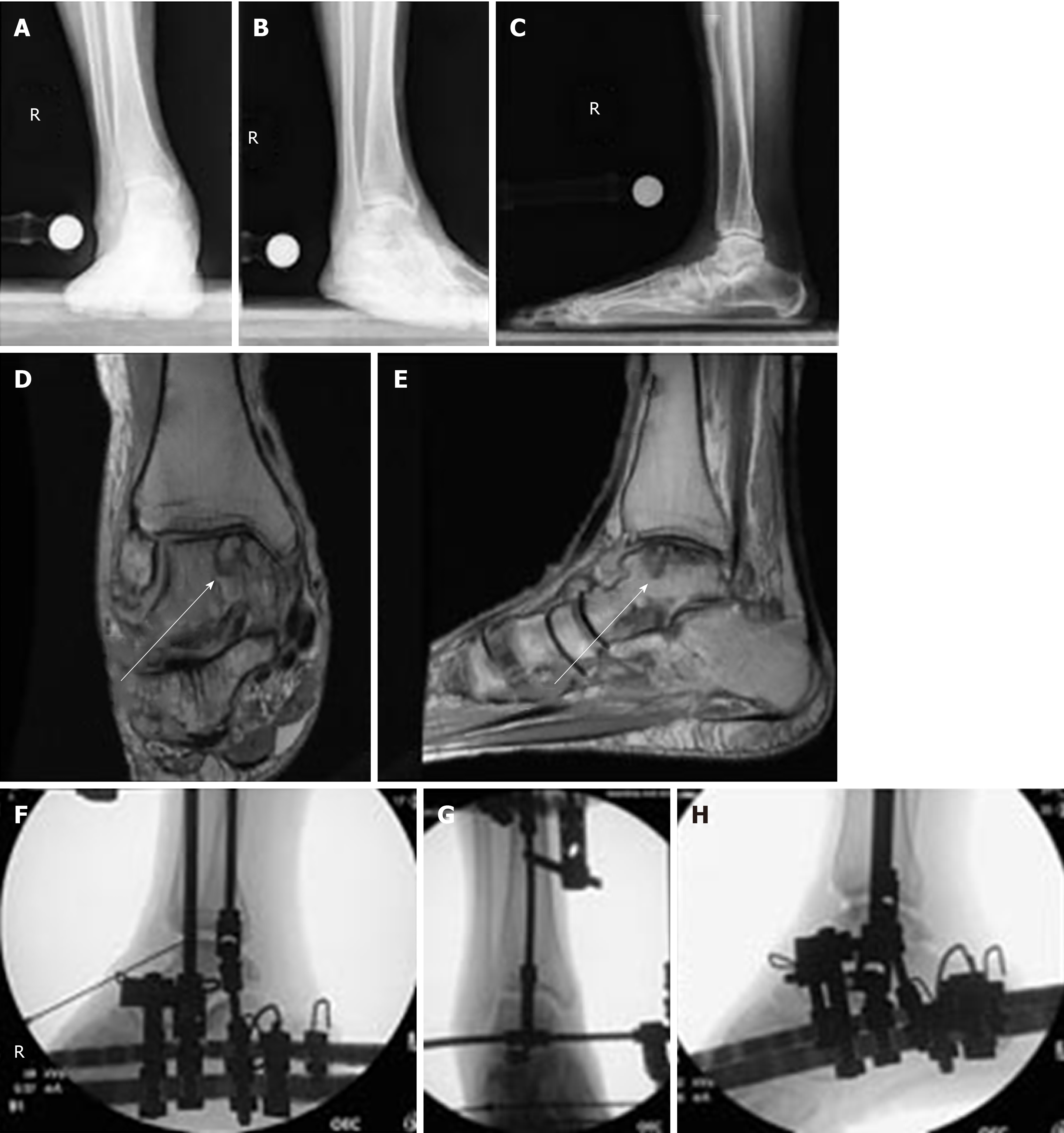
Figure 5 pre-operative standing X-rays and magnetic reconnaissance imaging, in addition to intra-operative fluoroscopy of the right ankle after anterior cheilectomy and distraction.
A: Anterior-Posterior (AP) X-rays demonstrates post-traumatic ankle osteoarthritis, joint space narrowing, anterior osteophyte, and flat foot deformity; B: Mortise view showing the same; C: Lateral view; D: Coronal view of the magnetic reconnaissance imaging shows medial osteochondritis dissecans lesion on the medial talar dome, as indicated by arrow; E: Sagittal view of the magnetic reconnaissance imaging showing medial osteochondritis dissecans lesion on the medial talar dome, as indicated by arrow; F: Anterior-Posterior intra-operative fluoroscopy of the right ankle after anterior cheilectomy, application of frame and distraction; G: Ankle in plantarflexion; H: Ankle in dorsiflexion with adjunctive injection of bone marrow aspirate concentrate into the ankle joint.
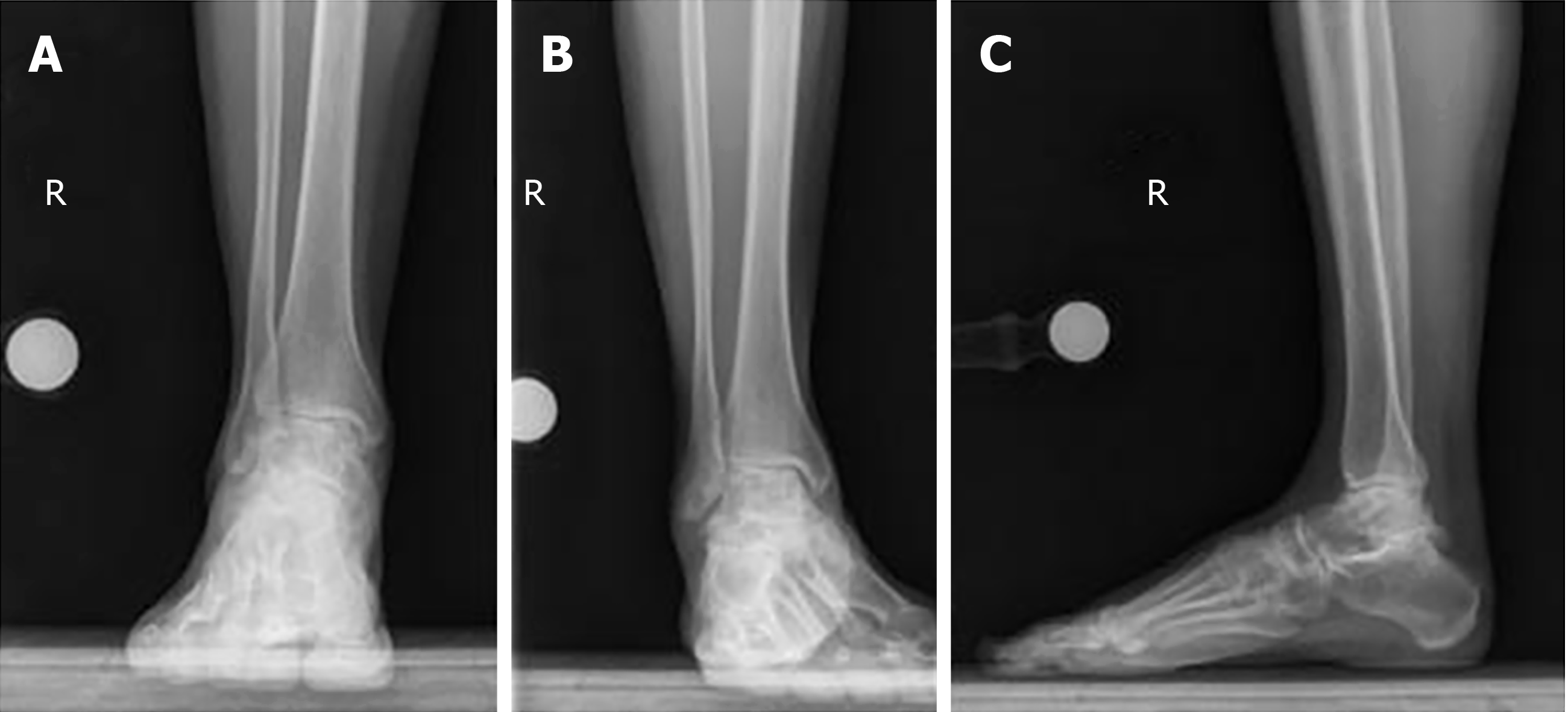
Figure 6 One-year post-operative standing X-rays of the right ankle, demonstrating improved ankle joint space, articular cartilage, and joint alignment one year after removal of frame.
A: Anterior-Posterior view; B: Mortise view; C: Lateral view.
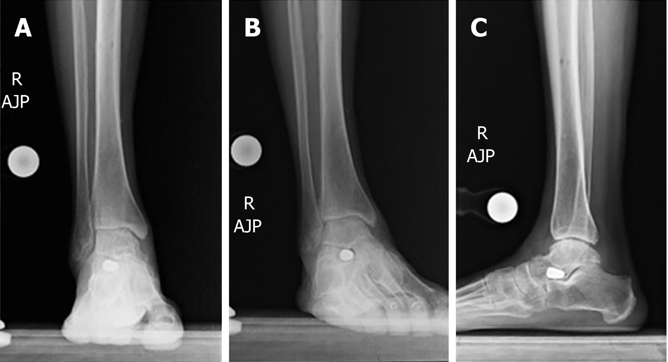
Figure 7 Pre-operative standing X-rays of the right subtalar joint demonstrating sub-talar joint space narrowing and post-traumatic osteoarthritis.
A: Anterior-Posterior view; B: Mortise view; C: Lateral view.
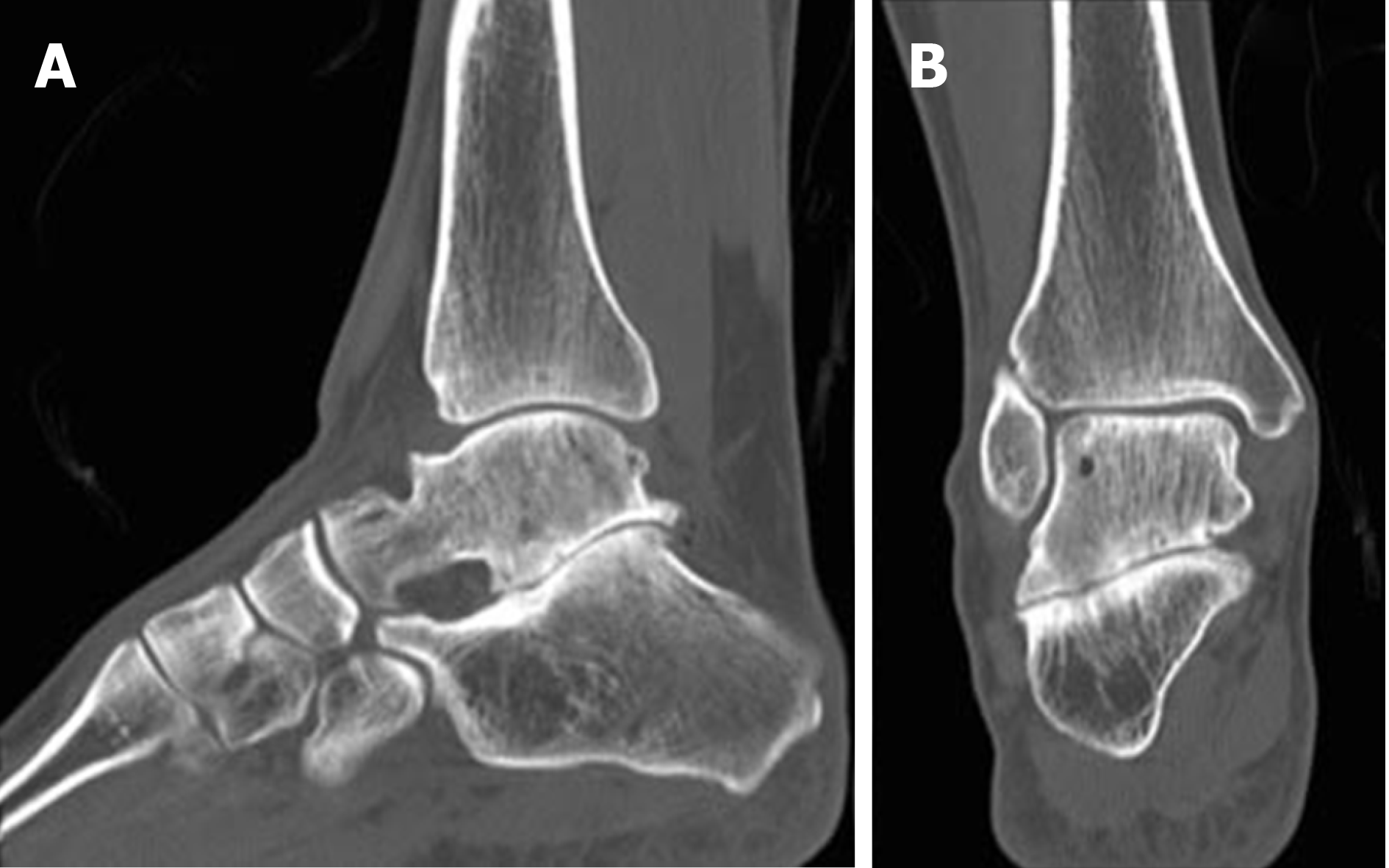
Figure 8 Pre-operative computed tomography scans of the right subtalar joint demonstrating sub-talar arthritis and sclerosis of the articular surface.
A: Sagittal view; B: Coronal view.
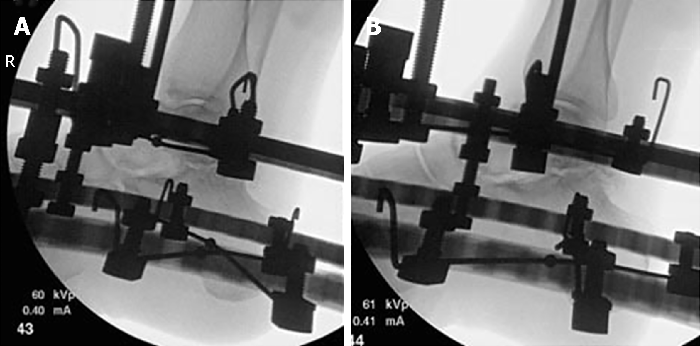
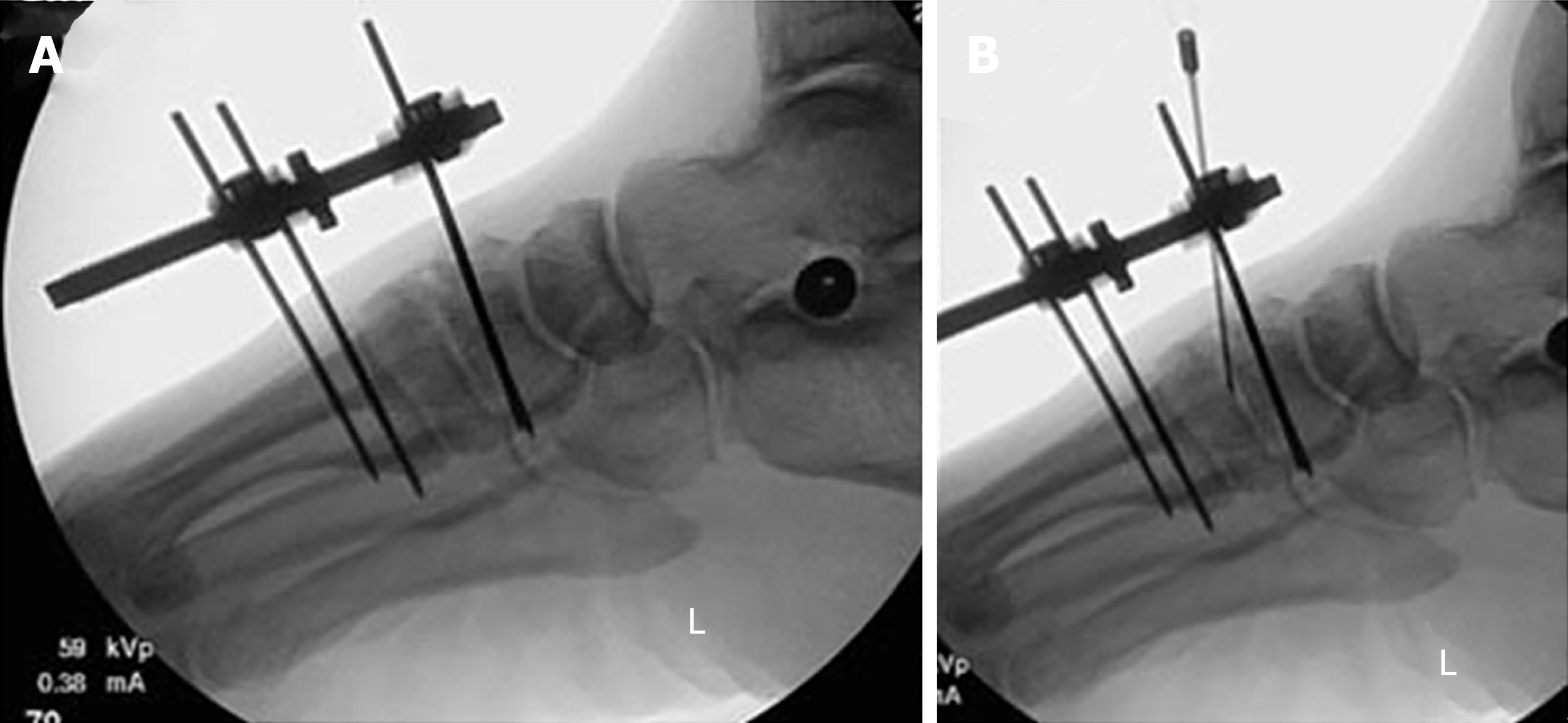
Figure 9 Intra-operative fluoroscopy of the right sub-talar joint before and after application of intraoperative distraction.
A: Lateral view before the distraction; B: Lateral view after 3 mm distraction.
Figure 10 One-month post-operative standing X-rays of the right sub-talar joint distraction, demonstrating widening of sub-talar joint space.
A: Anterior-Posterior view; B: Lateral view.
Figure 11 Comparison of pre and one-year post-operative standing X-rays of the right sub-talar joint.
A: Lateral view pre-operative standing X-ray; B: Lateral view post-operative standing X-ray one year after frame removal.
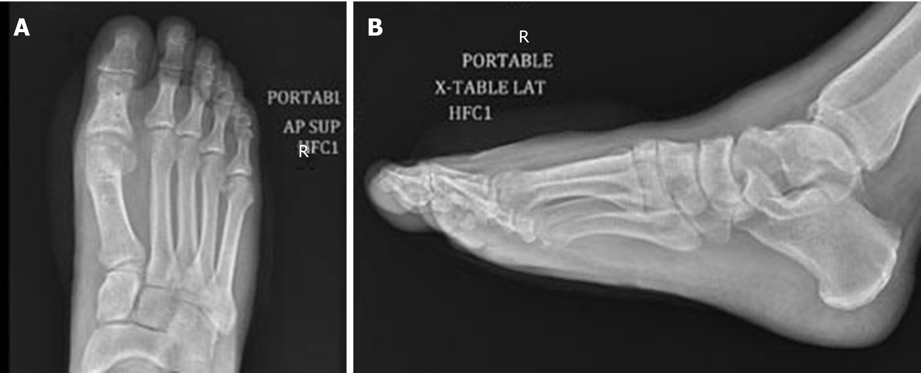
Figure 12 Pre-operative X-rays of the first metatarsophalangeal joint demonstrating hallux rigidus of the first metatarsophalangeal.
A: Anterior-Posterior view; B: Oblique view; C: Lateral view.
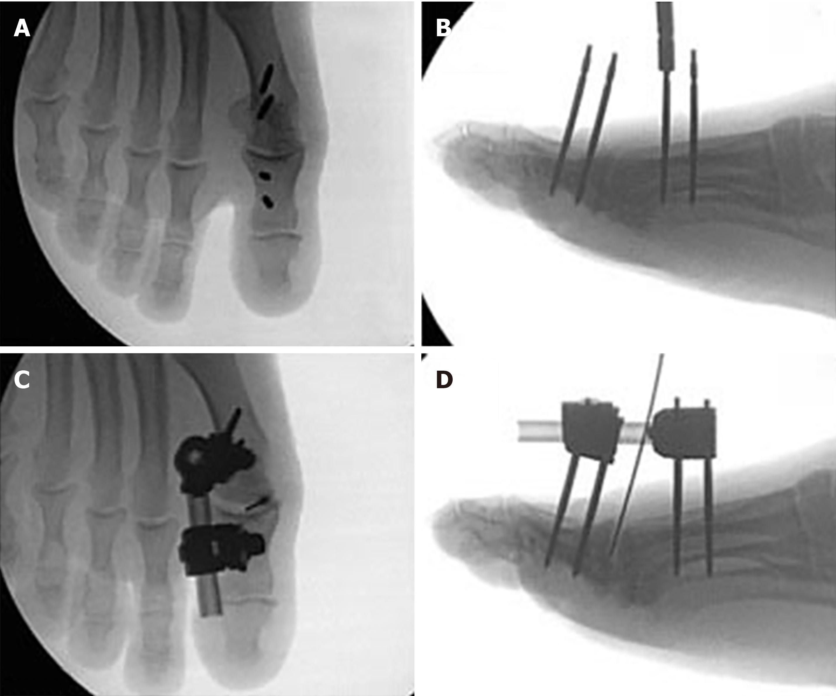
Figure 13 Intra-operative fluoroscopy of the right first metatarsophalangeal joint before and after application of external fixator.
A: Intra-operative fluoroscopy of the right first metatarsophalangeal (MTP) joint in the anterior-posterior view; B: Intra-operative fluoroscopy of the right first MTP joint in the view after insertion of pins; C: Intra-operative fluoroscopy of the right first MTP joint in the AP view after application of monolateral external fixator and injection of bone marrow aspirate concentrate; D: Intra-operative fluoroscopy of the right first MTP joint in lateral view after application of monolateral external fixator and injection of bone marrow aspirate concentrate. MTP: Metatarsophalangeal.
Figure 14 Three-month post-operative X-rays of the first metatarsophalangeal joint demonstrating improved metatarsophalangeal joint space.
A: Anterior-posterior view; B: Lateral view.
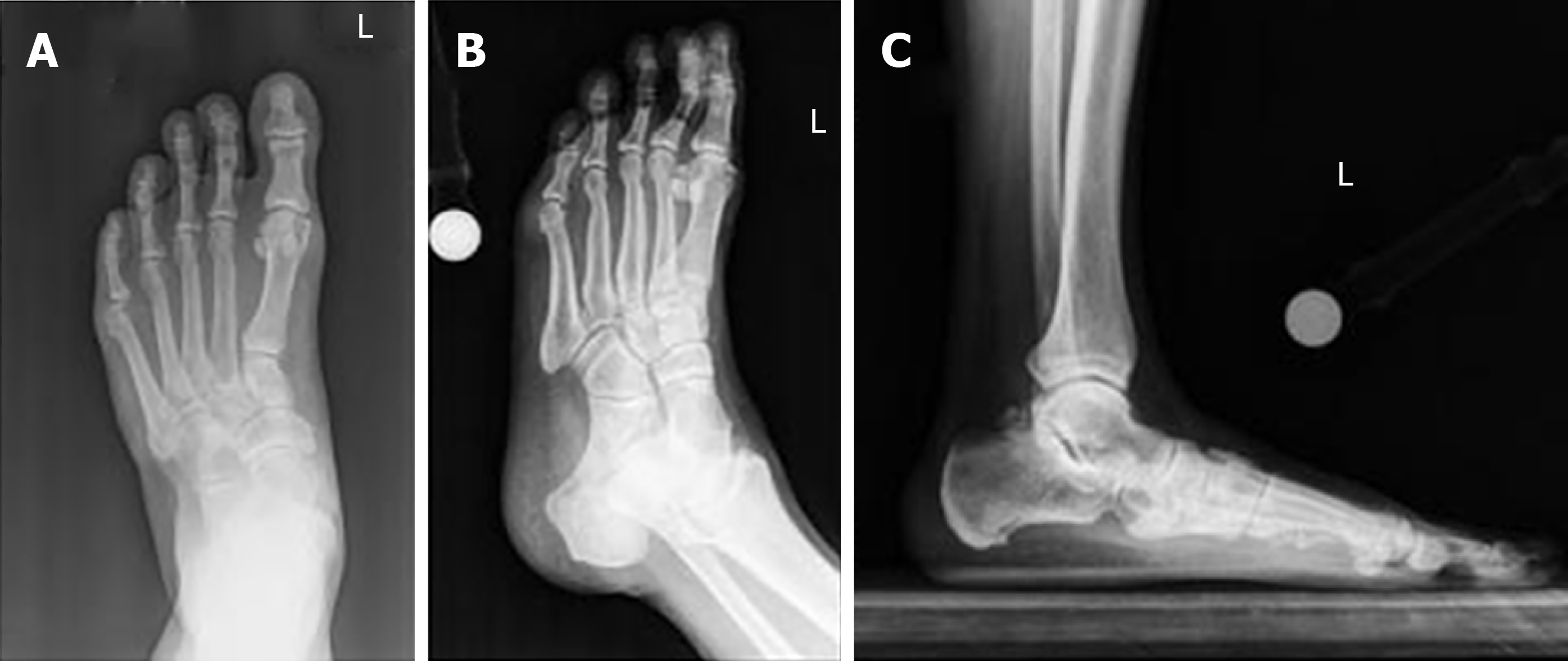
Figure 15 Pre-operative standing X-rays of the left second tarsometatarsal joint, demonstrating joint space narrowing and post-traumatic osteoarthritis in addition to flat foot deformity.
A: Anterior-posterior view; B: Oblique view; C: Lateral view.
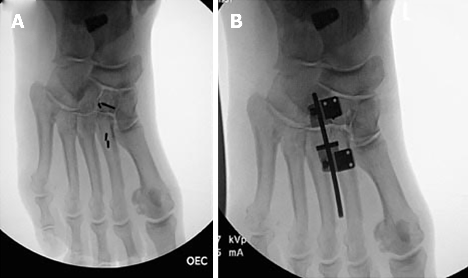
Figure 16 Intra-operative fluoroscopy of the left second tarsometatarsal joints demonstrating insertion of arthroeresis screw into the sub-talar joint to treat flat-foot deformity, in addition to application of external fixator.
A: Insertion of pins; B: Application of monolateral external fixator.
Figure 17 Intra-operative fluoroscopy of the left second tarsometatarsal joints joint demonstrating injection of bone marrow aspirate concentrate and application of external fixator.
A: Lateral view after application of frame; B: Bone marrow aspirate concentrate.
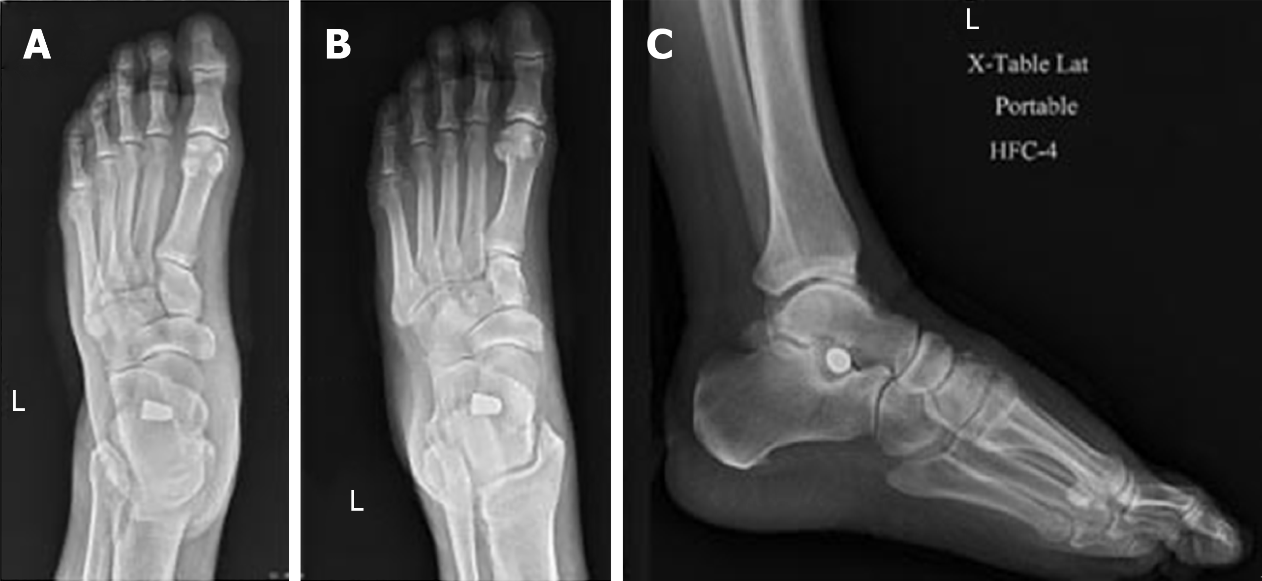
Figure 18 Three-months post-operative X-rays of the left second tarsometatarsal joint, demonstrating improved tarsometatarsal joint space and articular cartilage.
A: Anterior-posterior view; B: Oblique view; C: Lateral view.
- Citation: Dabash S, Buksbaum JR, Fragomen A, Rozbruch SR. Distraction arthroplasty in osteoarthritis of the foot and ankle. World J Orthop 2020; 11(3): 145-157
- URL: https://www.wjgnet.com/2218-5836/full/v11/i3/145.htm
- DOI: https://dx.doi.org/10.5312/wjo.v11.i3.145


