Copyright
©The Author(s) 2020.
World J Orthop. Feb 18, 2020; 11(2): 129-136
Published online Feb 18, 2020. doi: 10.5312/wjo.v11.i2.129
Published online Feb 18, 2020. doi: 10.5312/wjo.v11.i2.129
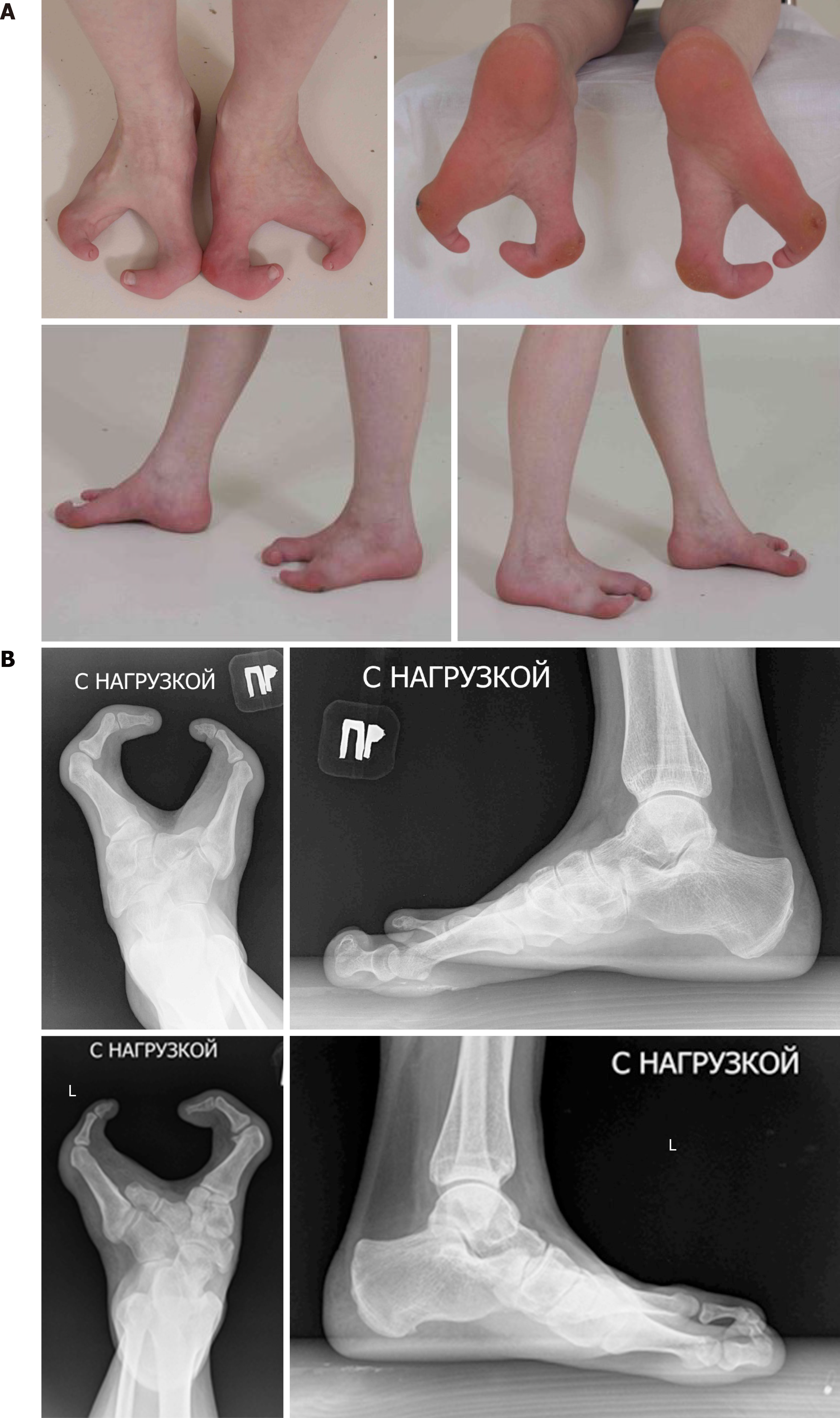
Figure 1 Photo and x-ray pictures of patient’s feet before treatment.
A: Cleft feet; B: X-rays of feet in anterior-posterior and lateral view (absence of central feet rays).
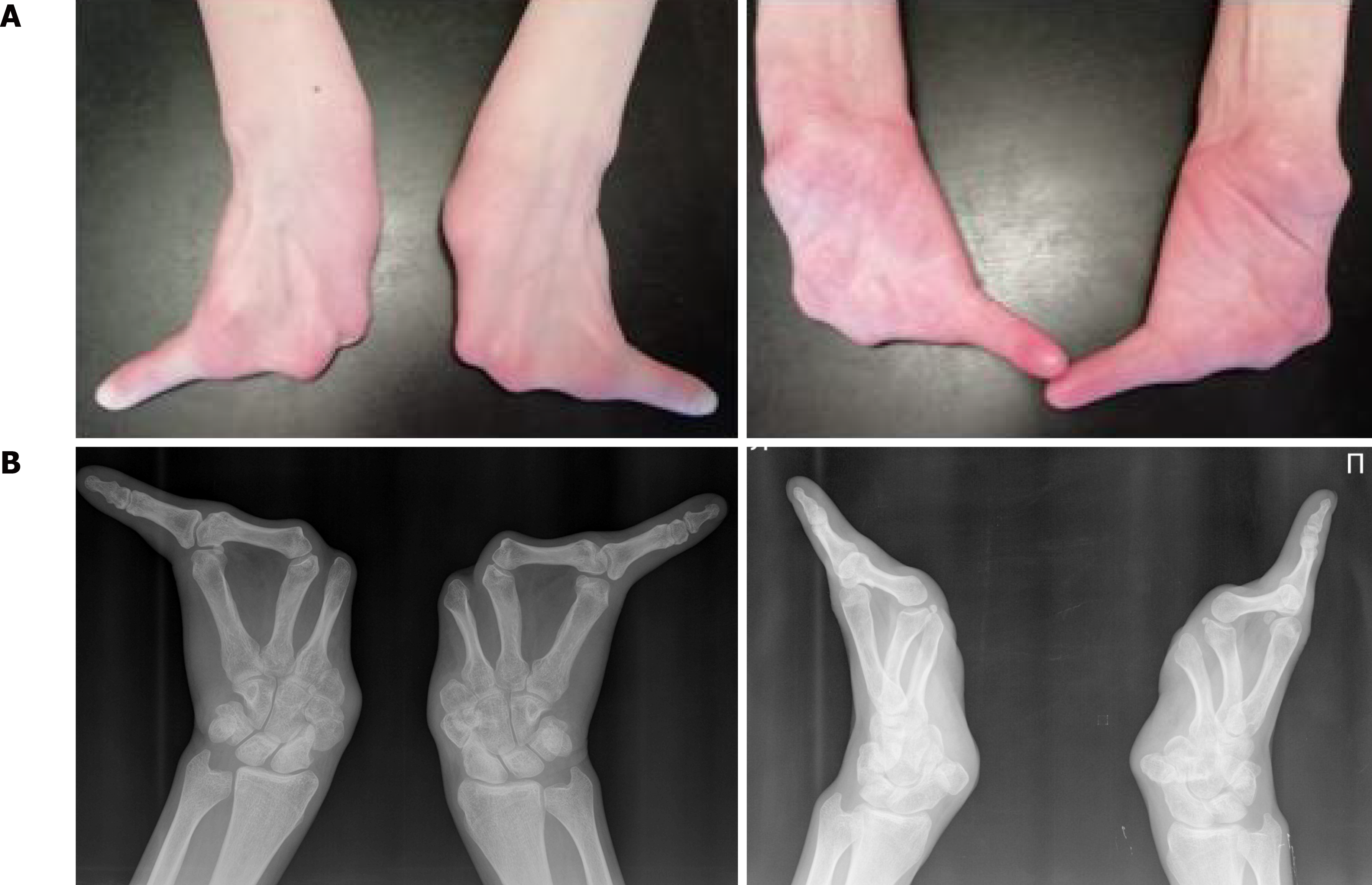
Figure 2 Photo and x-ray pictures of patient’s hands.
A: Split hands with absence of fingers 1-4; B: Three metacarpals with transverse bone in base of cleft and absence of fingers 1-4.
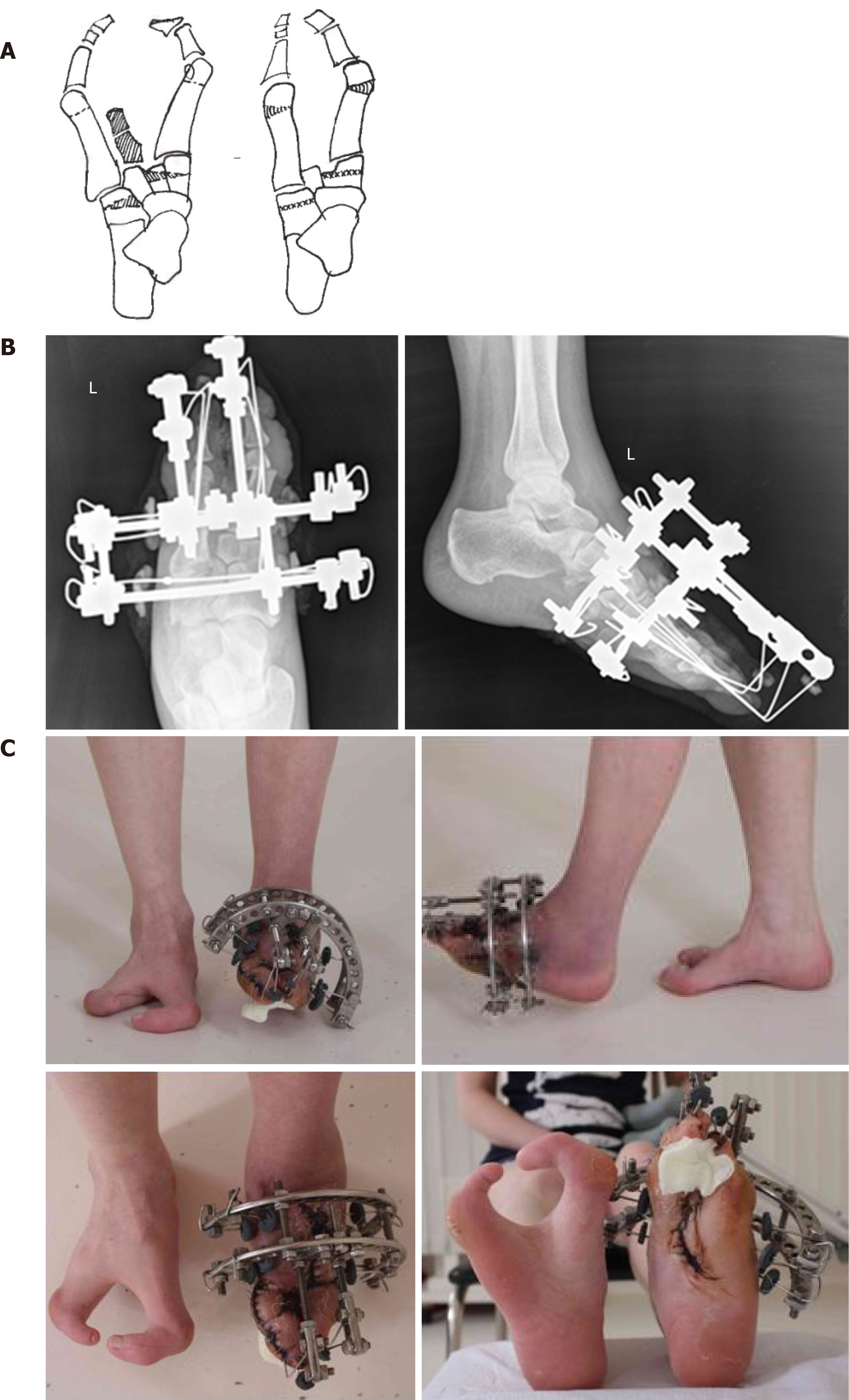
Figure 3 Scheme of surgical intervention, x-rays of left foot and photo of feet during treatment process.
A: The first step was resectional wedge-shaped osteotomy of cuboid and cuneiform bones with removing of rudiment of central foot ray. The second step was corrective osteotomy of both metatarsal bones; B: Correction and fixation of foot rays by minimalist construct of Ilizarov apparatus; C: Closure of foot splitting.
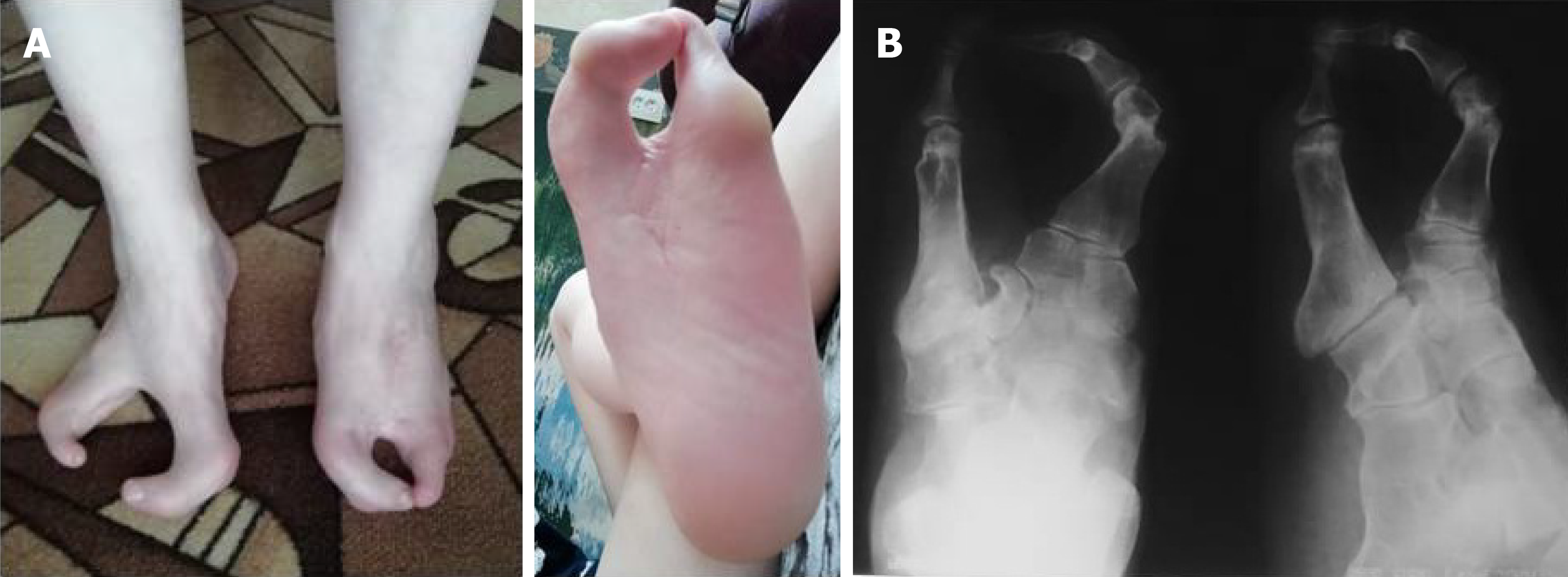
Figure 4 Photo of feet and x-ray pictures of patient’s left foot after 2 years after surgical intervention.
A: Closure of foot splitting; B: X-rays of left foot in anterior-posterior and axial view.
- Citation: Leonchuk SS, Neretin AS, Blanchard AJ. Cleft foot: A case report and review of literature. World J Orthop 2020; 11(2): 129-136
- URL: https://www.wjgnet.com/2218-5836/full/v11/i2/129.htm
- DOI: https://dx.doi.org/10.5312/wjo.v11.i2.129


