Copyright
©The Author(s) 2020.
World J Orthop. Nov 18, 2020; 11(11): 523-527
Published online Nov 18, 2020. doi: 10.5312/wjo.v11.i11.523
Published online Nov 18, 2020. doi: 10.5312/wjo.v11.i11.523
Figure 1 Preoperative examinations of the patient.
A: Preoperative physical examination showed many café-au-lait spots and severe spinal deformity; B: An axial computerized tomography image at L1-L5 demonstrates the thin pedicles; C: Axial T2-W images obtained by magnetic resonance imaging show vertebral scalloping with a dural ectasia; D: A three-dimensional model shows the preoperative vertebral condition.
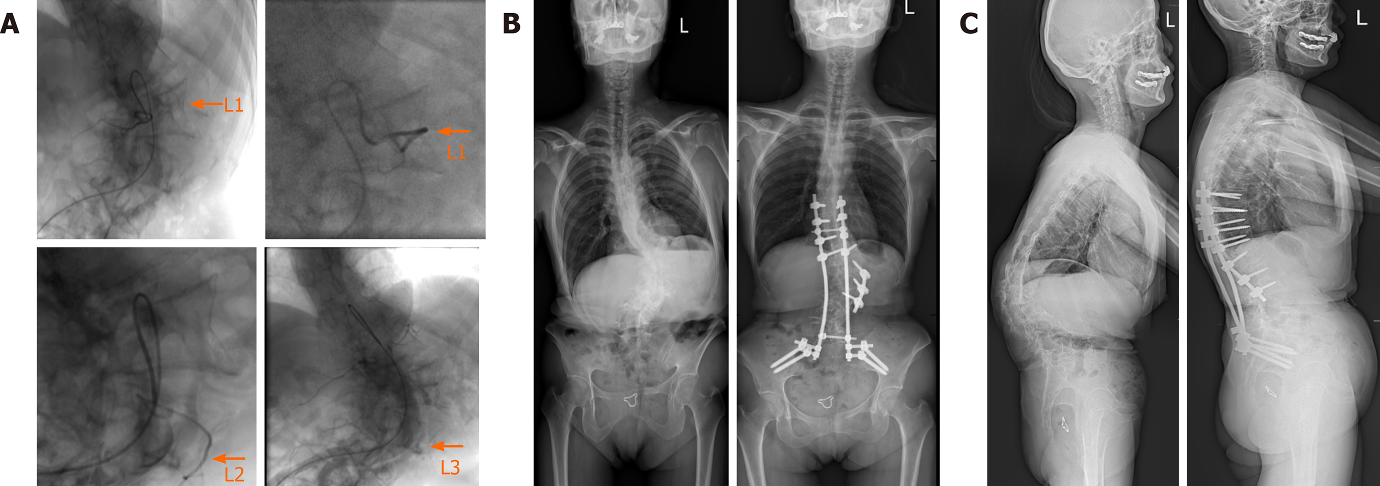
Figure 2 Images of the patient before and after the surgery.
A: Digital subtraction angiography images show the lumbar vascular conditions at 48 h before the surgery; B and C: Plain radiographs at the two-year follow-up indicate the successful correction (right) of the spinal deformity, when compared to the preoperative ones (left).
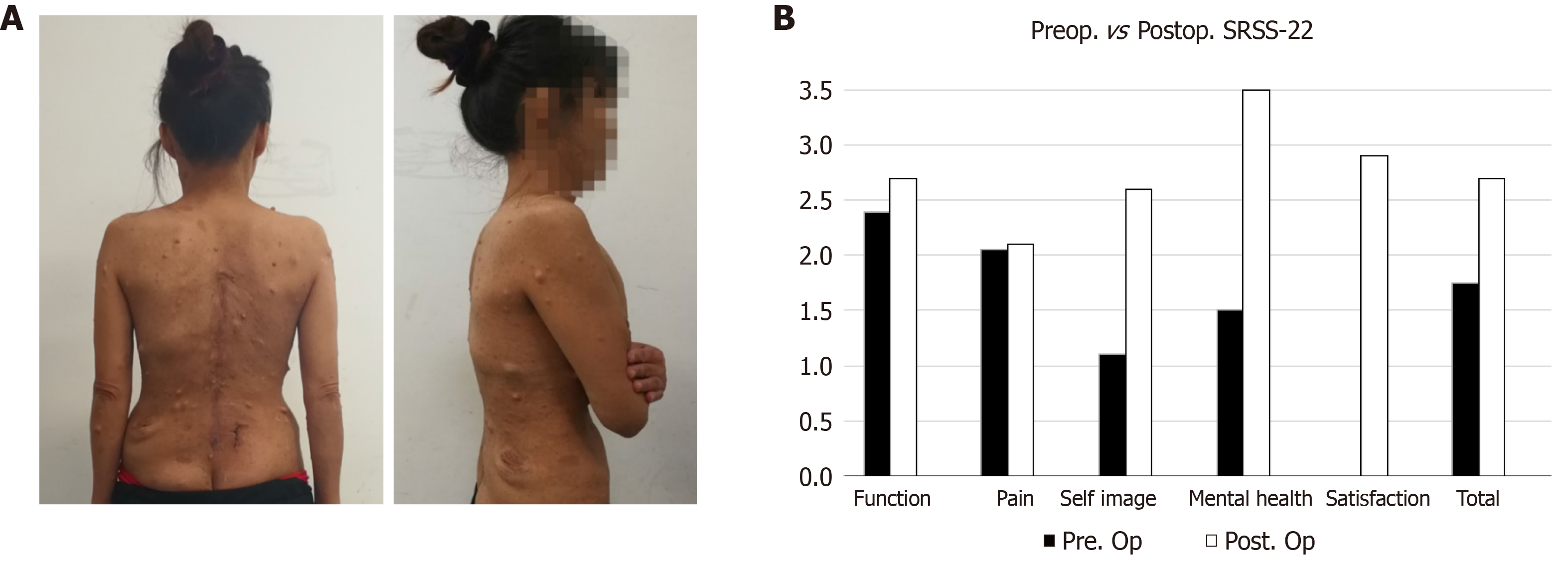
Figure 3 Patient photos and the Scoliosis Research Society-22r score at the two-year follow-up.
A: The deformity had been improved obviously; B: Scoliosis Research Society-22r score indicates a significant improvement after surgery. Pre. Op: Preoperative; Post. Op: Postoperative.
- Citation: Shi Y, Li YH, Guan ZP, Huang YC, Yu BS. Modified surgical treatment for a patient with neurofibromatosis scoliosis: A case report. World J Orthop 2020; 11(11): 523-527
- URL: https://www.wjgnet.com/2218-5836/full/v11/i11/523.htm
- DOI: https://dx.doi.org/10.5312/wjo.v11.i11.523


