Copyright
©The Author(s) 2020.
World J Orthop. Nov 18, 2020; 11(11): 507-515
Published online Nov 18, 2020. doi: 10.5312/wjo.v11.i11.507
Published online Nov 18, 2020. doi: 10.5312/wjo.v11.i11.507
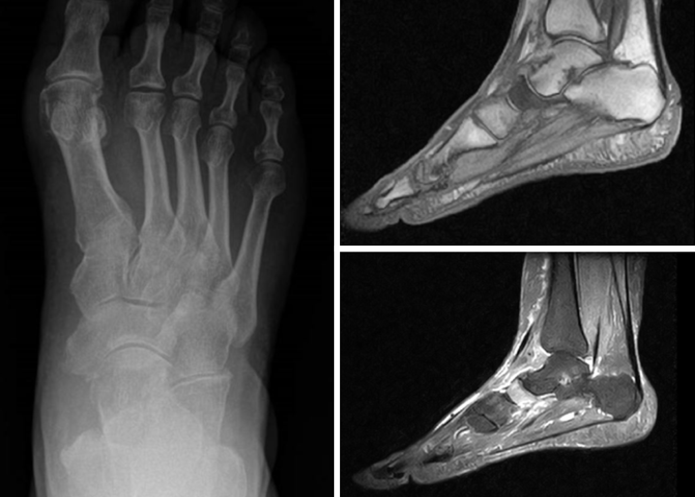
Figure 1 Typical tarsal “scaphoiditis” of Müller-Weiss disease is shown: Early arthritis in X-rays and signal alterations in magnetic resonance imaging.
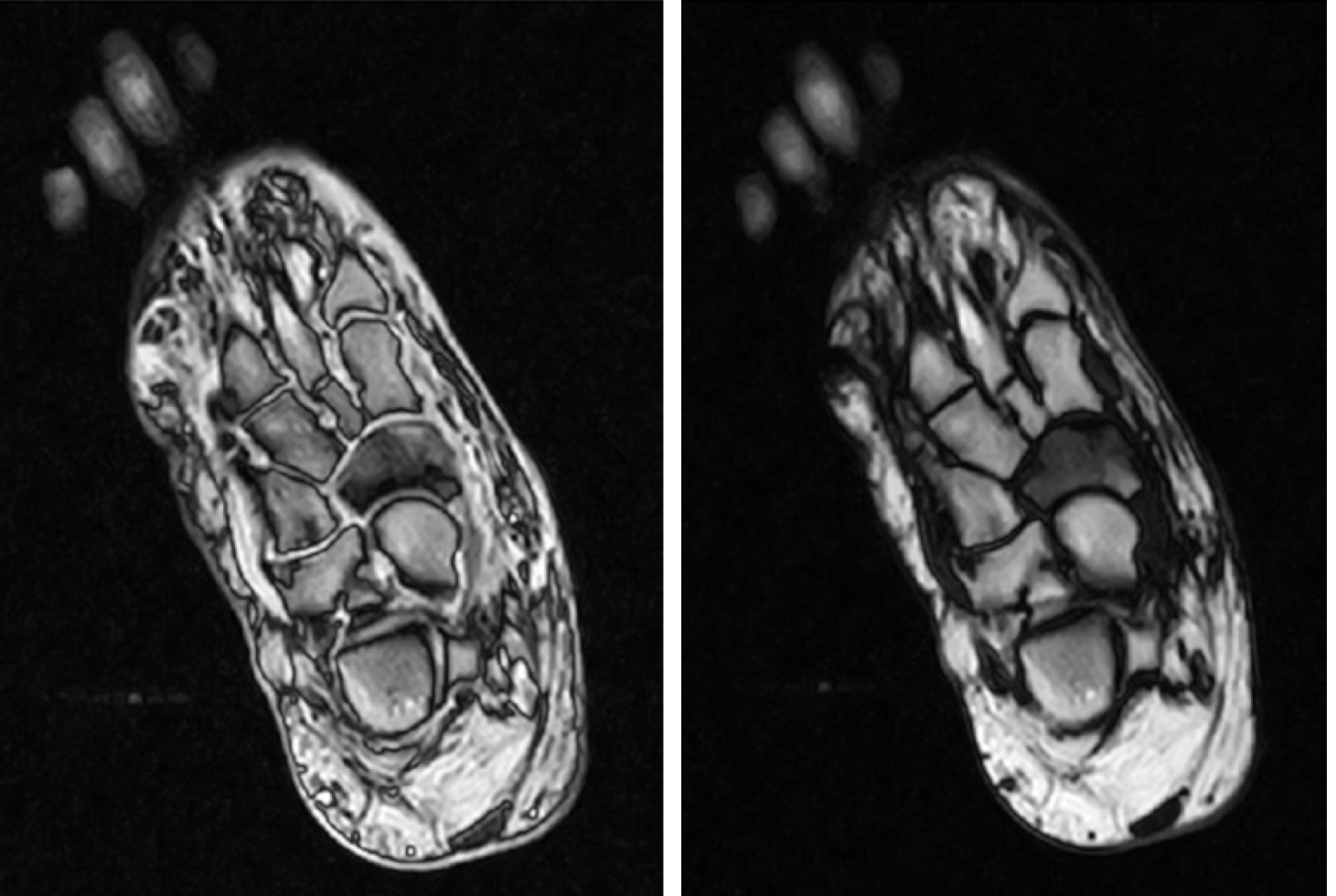
Figure 2 Magnetic resonance imaging reveals signal alterations and fragmentation of the navicular in Müller-Weiss disease.
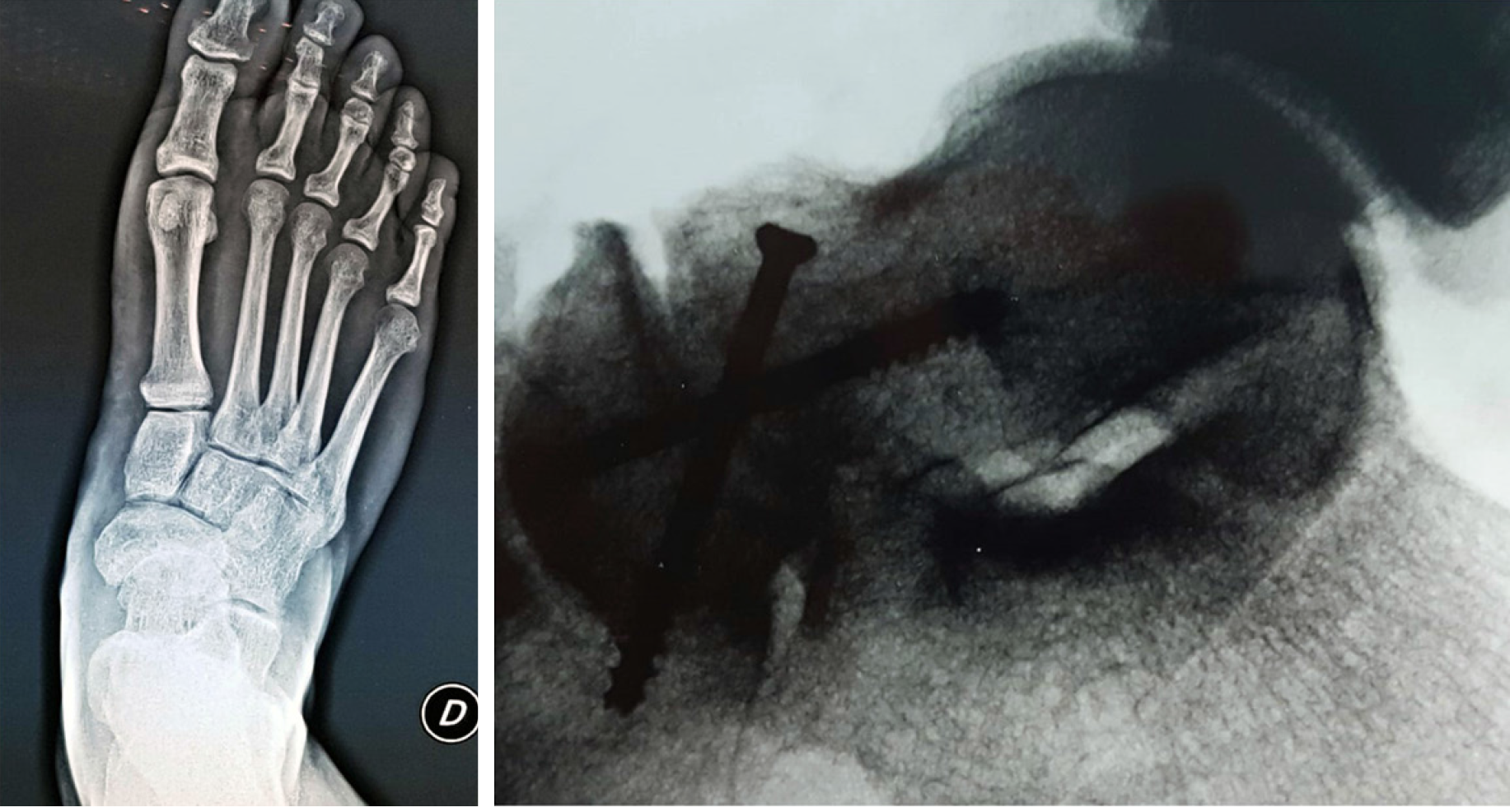
Figure 3 Arthritis of the talo-navicular joint is shown in X-rays: An arthrodesis with two screws was performed.
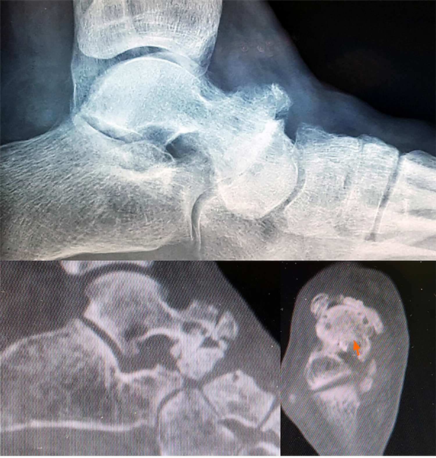
Figure 4 X-rays and computed tomography scans show advanced arthritis of the talo-navicular joint.
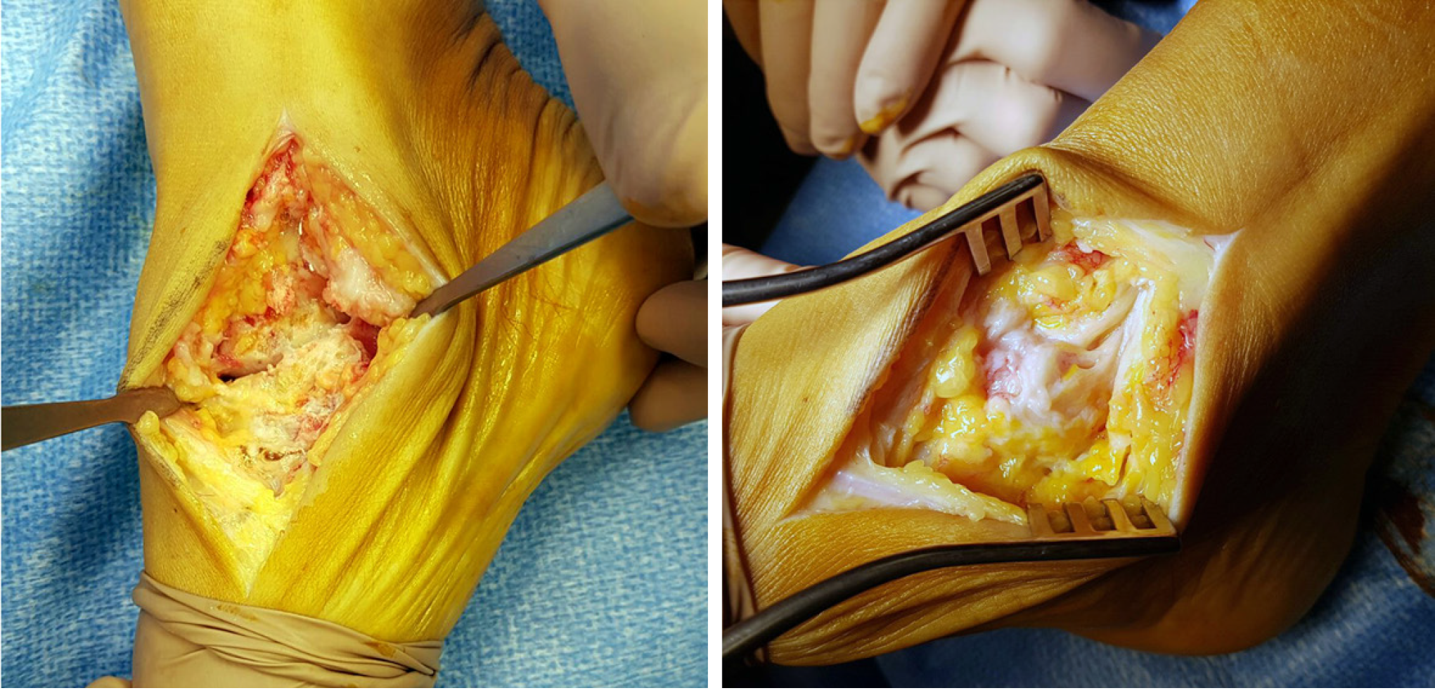
Figure 5 Patient underwent talo-navicular arthrodesis.
Intra-operative images show degeneration of the joint.
- Citation: Volpe A, Monestier L, Malara T, Riva G, La Barbera G, Surace MF. Müller-Weiss disease: Four case reports. World J Orthop 2020; 11(11): 507-515
- URL: https://www.wjgnet.com/2218-5836/full/v11/i11/507.htm
- DOI: https://dx.doi.org/10.5312/wjo.v11.i11.507


