Copyright
©The Author(s) 2020.
World J Orthop. Oct 18, 2020; 11(10): 465-472
Published online Oct 18, 2020. doi: 10.5312/wjo.v11.i10.465
Published online Oct 18, 2020. doi: 10.5312/wjo.v11.i10.465
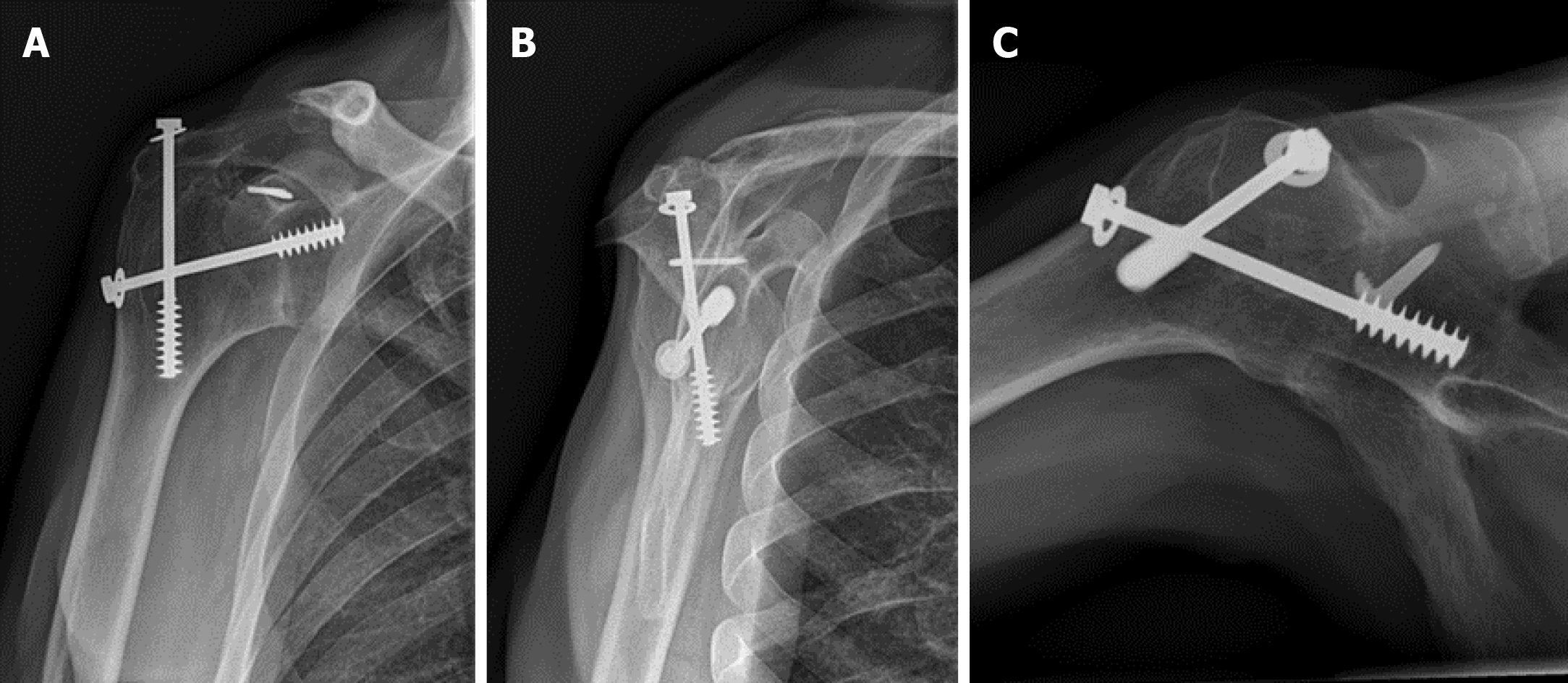
Figure 1 Conventional radiographs demonstrating the glenohumeral and acromiohumeral consolidation.
A: Preoperative anteroposterior; B: Lateral/scapular Y; C: Axial radiographs.
Figure 2 Preoperative Computed Tomography scan demonstrating the glenohumeral and acromiohumeral consolidation.
A: Coronal view; B: Sagittal view; C: Axial view.
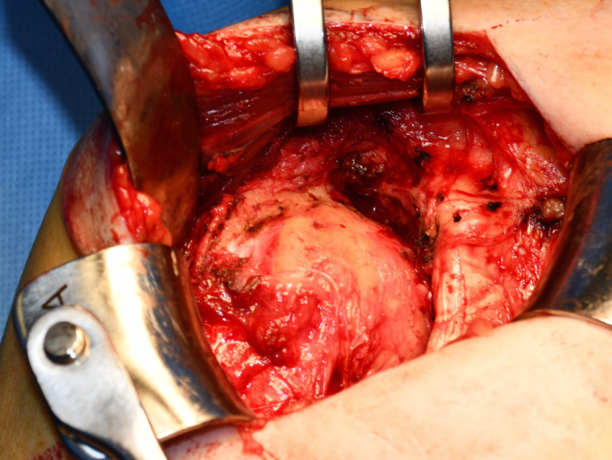
Figure 3 Deltopectoral approach.
The anterior and cranial rotator cuff muscles are absent.
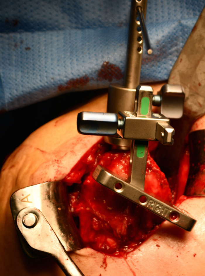
Figure 4 The glenohumeral osteotomy.
The glenohumeral osteotomy was performed under 10º of inclination, a neutral version and 1 cm lateral from the original joint line.
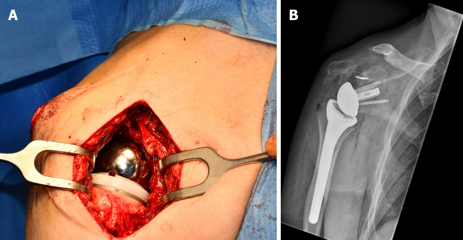
Figure 5 The implanted reverse shoulder arthroplasty.
A: During surgery; B: Postoperative anteroposterior radiograph.
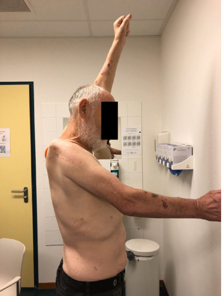
Figure 6 The active forward elevation of 50º after 12 mo postoperatively.
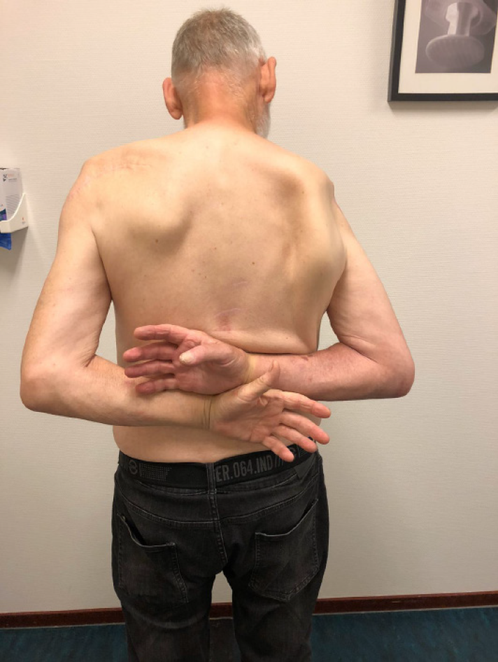
Figure 7 The internal rotation toTh12 12 mo postoperatively.
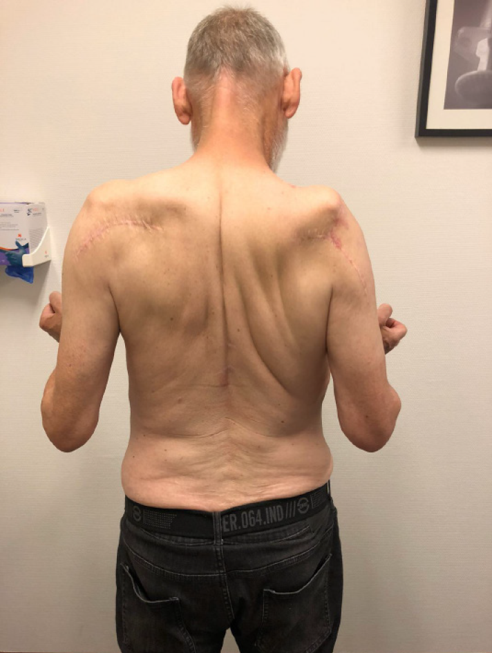
Figure 8 No external rotation 12 mo postoperatively.
- Citation: Dogger MN, Bemmel AFV, Alta TDW, van Noort A. Conversion to reverse shoulder arthroplasty fifty-one years after shoulder arthrodesis: A case report. World J Orthop 2020; 11(10): 465-472
- URL: https://www.wjgnet.com/2218-5836/full/v11/i10/465.htm
- DOI: https://dx.doi.org/10.5312/wjo.v11.i10.465


