Copyright
©The Author(s) 2019.
World J Orthop. Feb 18, 2019; 10(2): 71-80
Published online Feb 18, 2019. doi: 10.5312/wjo.v10.i2.71
Published online Feb 18, 2019. doi: 10.5312/wjo.v10.i2.71
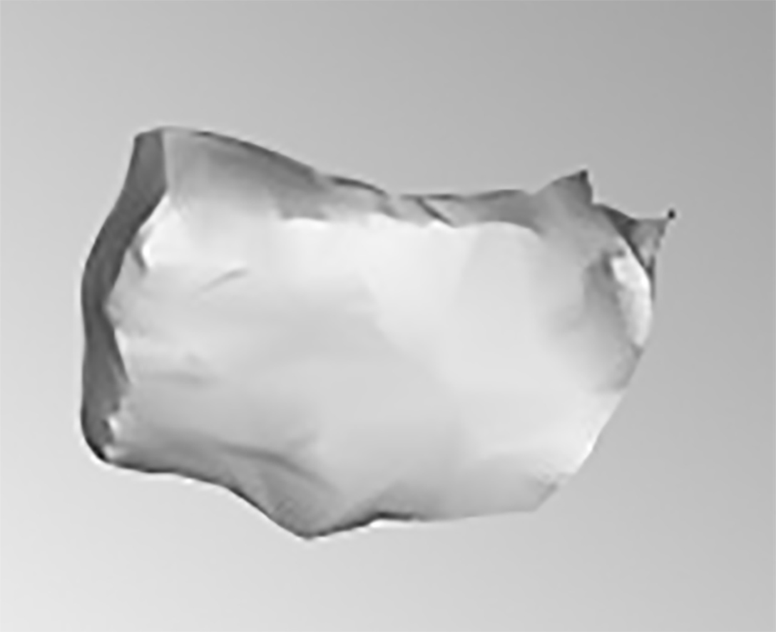
Figure 1 Cuboid bone lateral view.
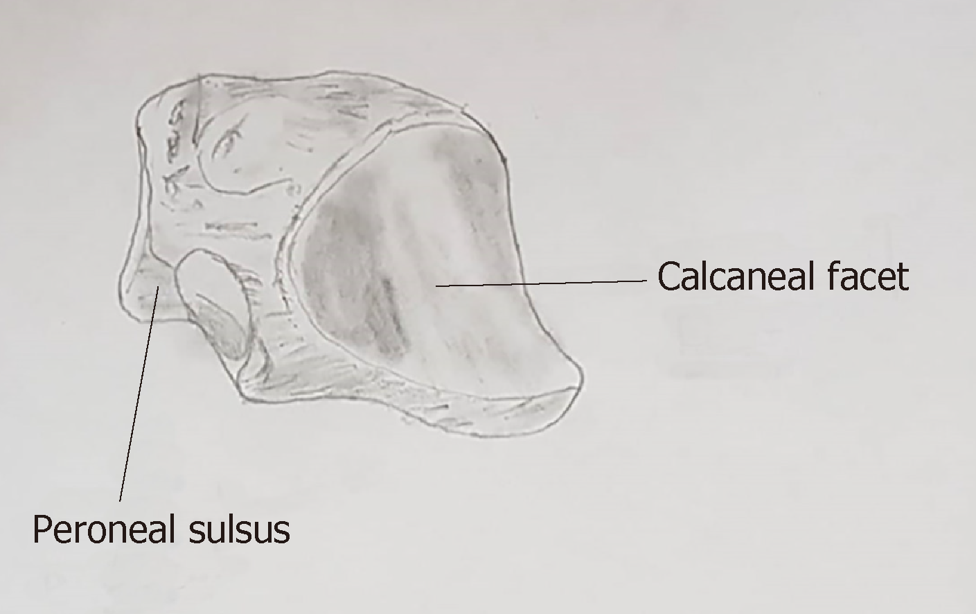
Figure 2 Posterolateral view of cuboid depicting peroneal groove (modified from Greys Anatomy, 1918).
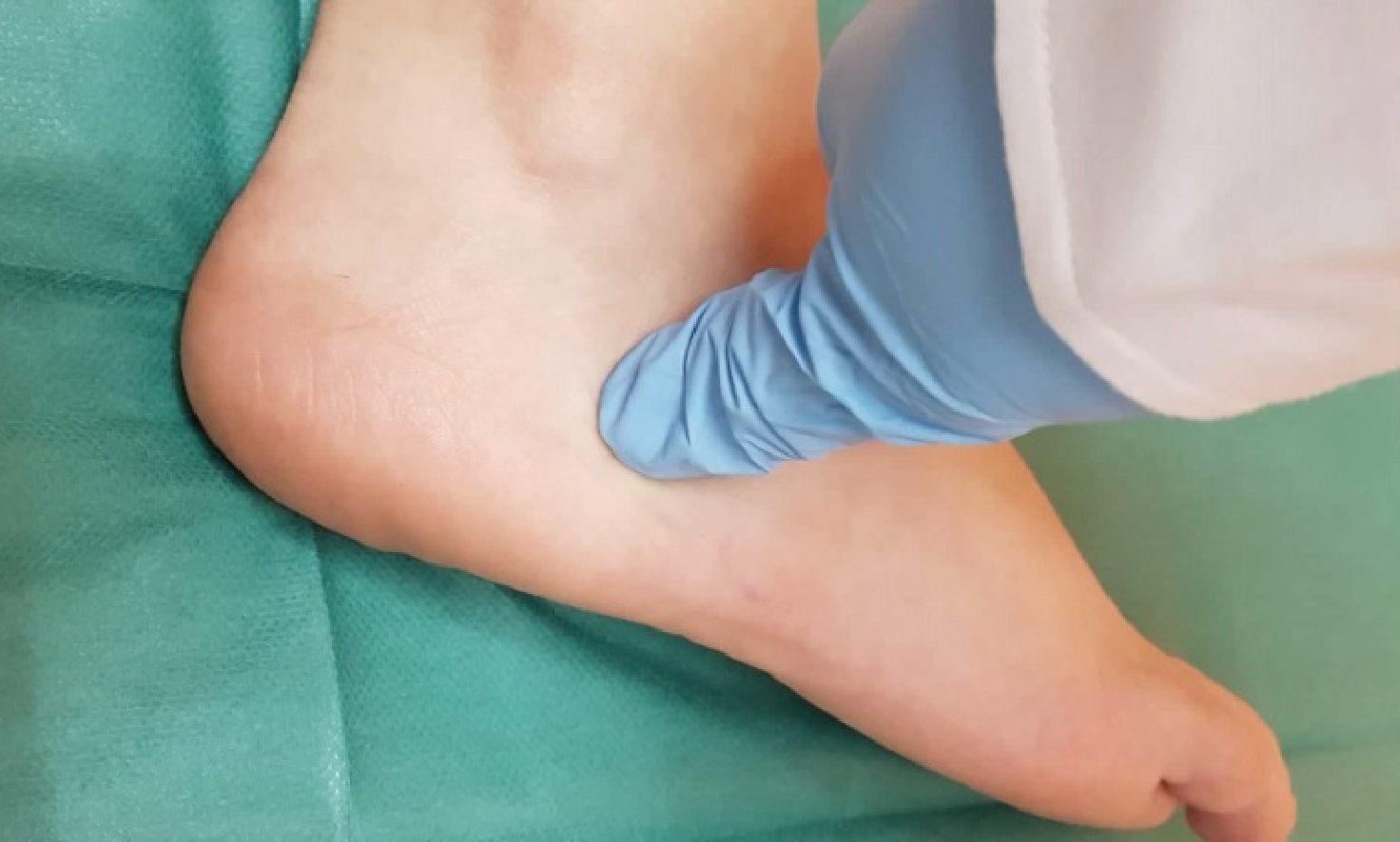
Figure 3 Local tenderness to direct palpation of the cuboid bone following foot injury may suggest cuboid fracture.
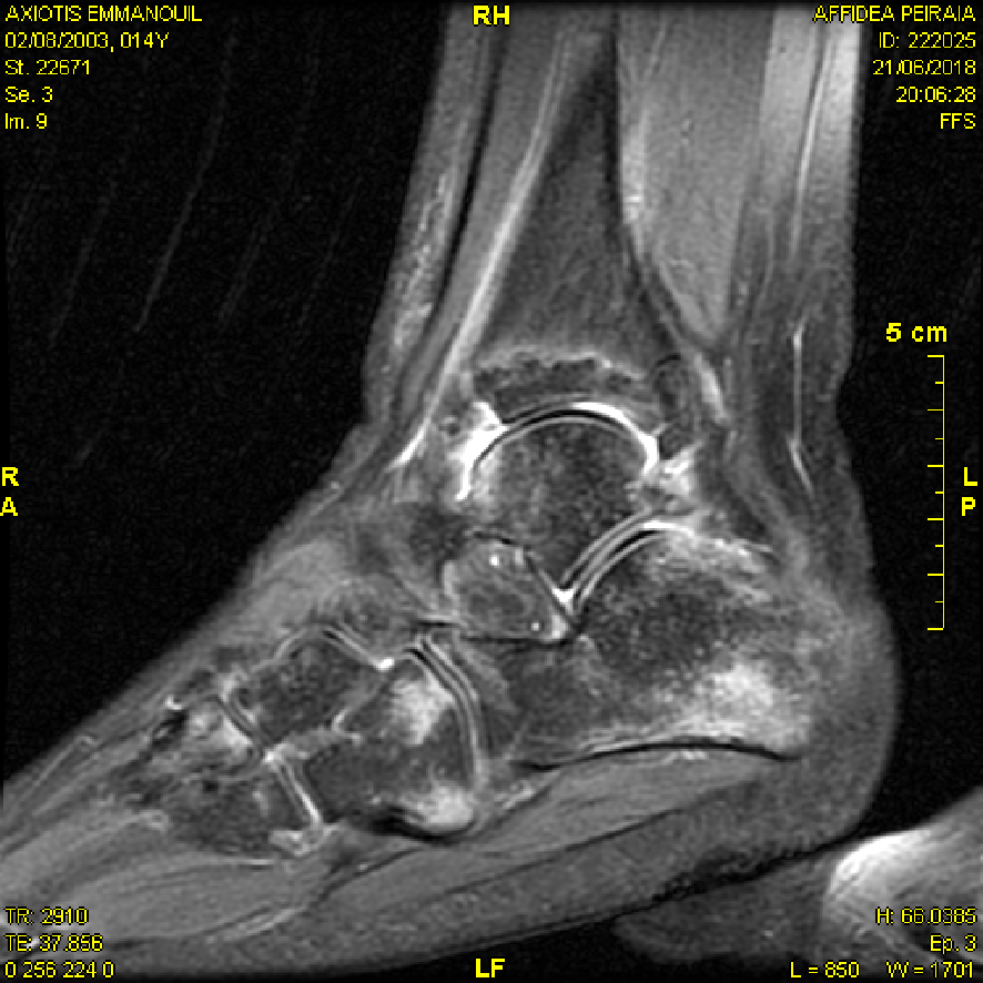
Figure 4 Sagittal view of the right ankle obtained via magnetic resonance imaging.
Undisplaced cuboid fracture extending to the middle of the calcaneocuboid joint.
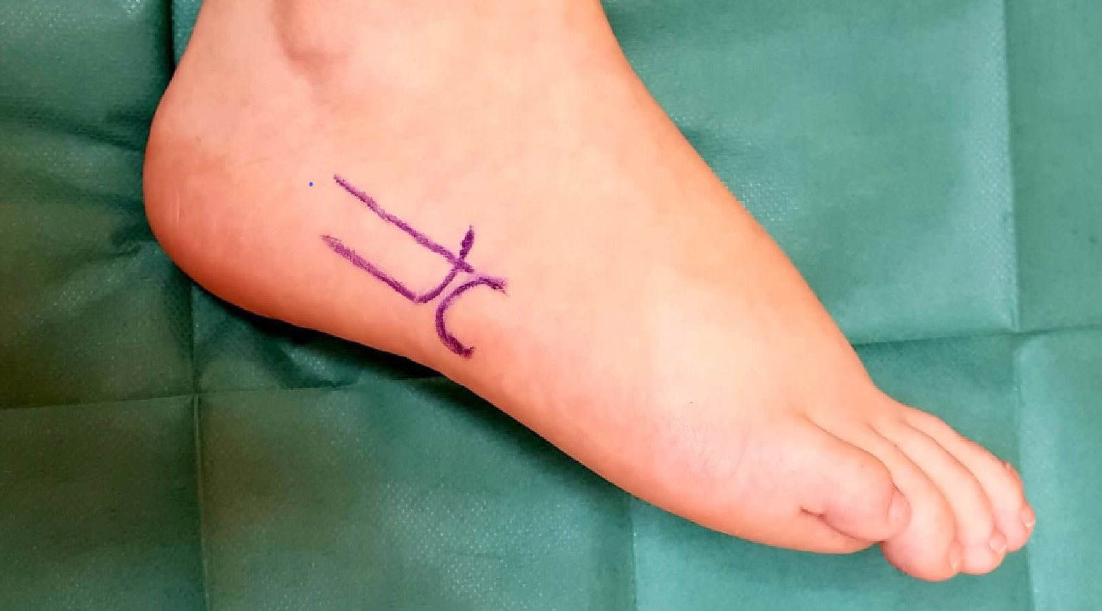
Figure 5 The lateral longitudinal surgical incision for the internal fixation of cuboid fractures.
- Citation: Angoules AG, Angoules NA, Georgoudis M, Kapetanakis S. Update on diagnosis and management of cuboid fractures. World J Orthop 2019; 10(2): 71-80
- URL: https://www.wjgnet.com/2218-5836/full/v10/i2/71.htm
- DOI: https://dx.doi.org/10.5312/wjo.v10.i2.71


