Copyright
©The Author(s) 2019.
World J Orthop. Nov 18, 2019; 10(11): 404-415
Published online Nov 18, 2019. doi: 10.5312/wjo.v10.i11.404
Published online Nov 18, 2019. doi: 10.5312/wjo.v10.i11.404
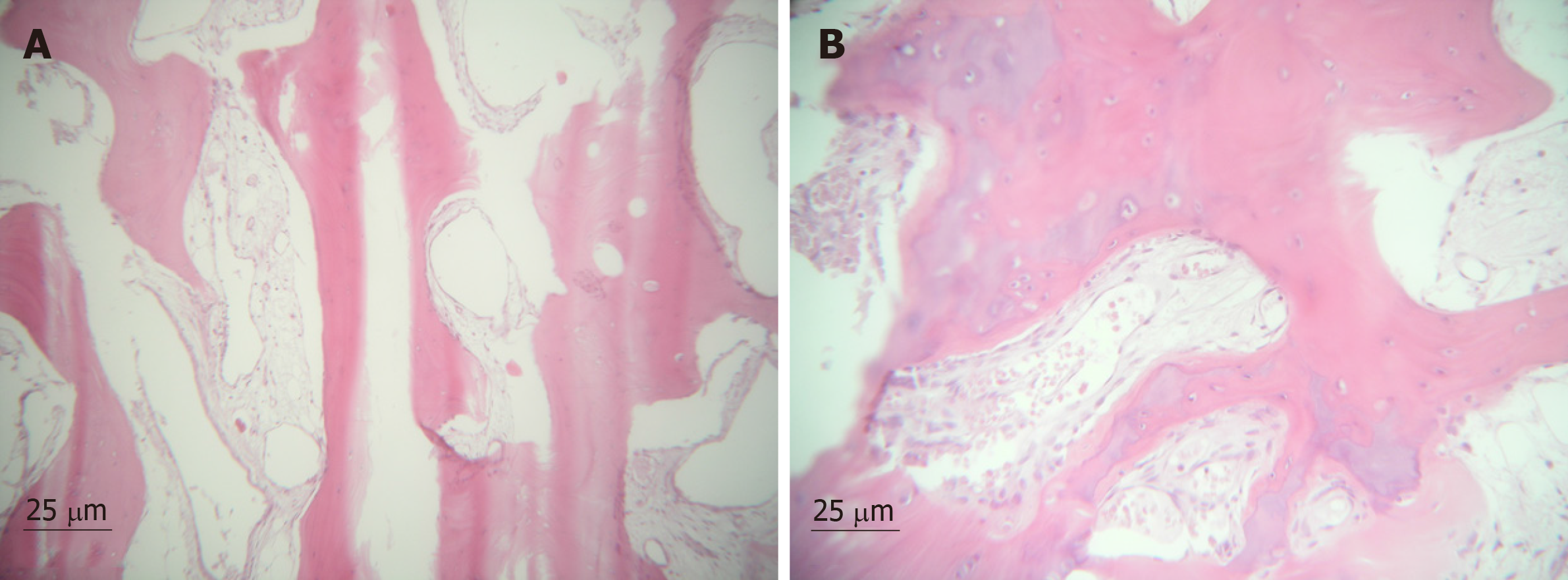
Figure 1 Histological features of synovium in chondromatosis: Trabecular and stroma, with collagen and new bone tissue forming; 10 × zoom (A) and 25 × zoom (B).
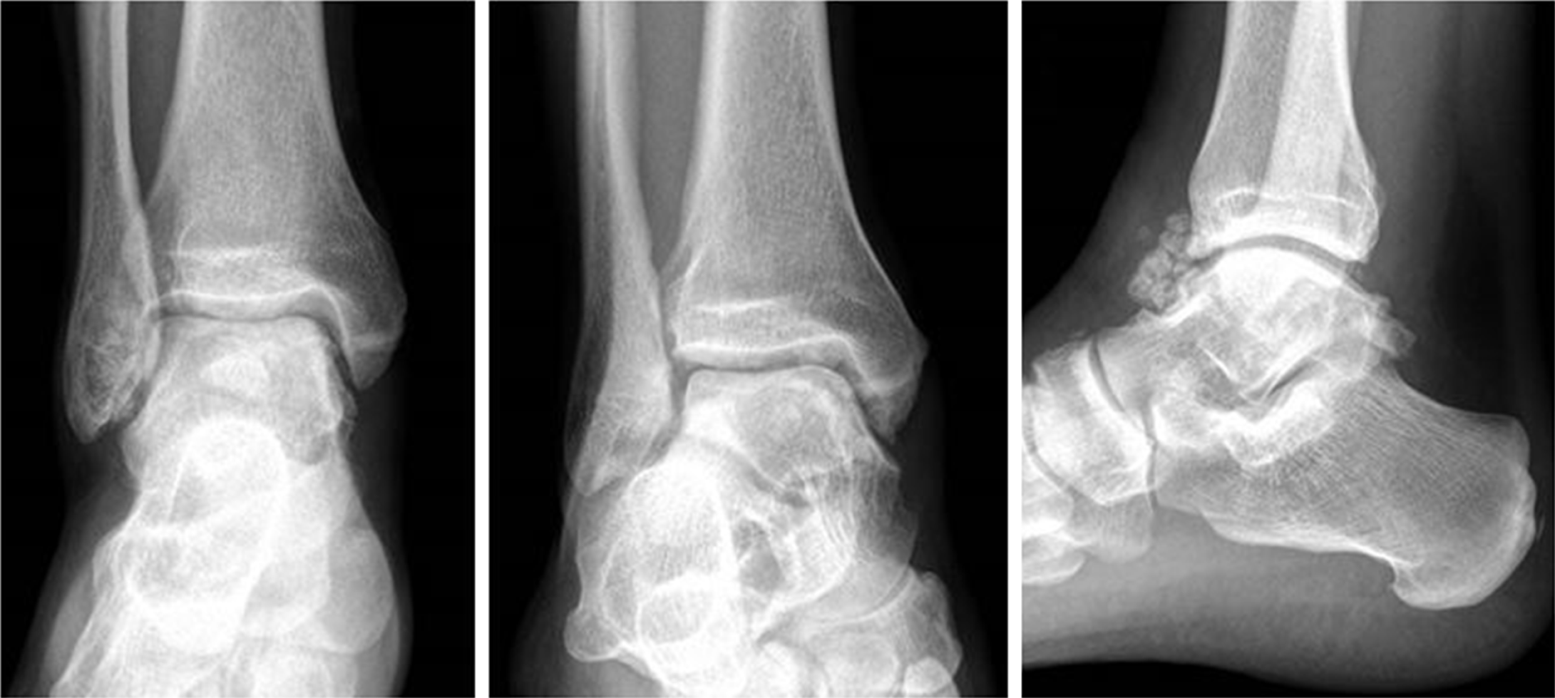
Figure 2 Radiographs of the ankle in Patient 1, after operative removal.
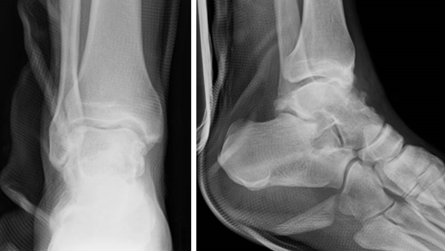
Figure 3 Pre-operative radiographs of Patient 2 with subtalar primary synovial chondromatosis.
Figure 4 Radiographs of the ankle in Patient 1, showing several loose bodies at the anterior compartment.
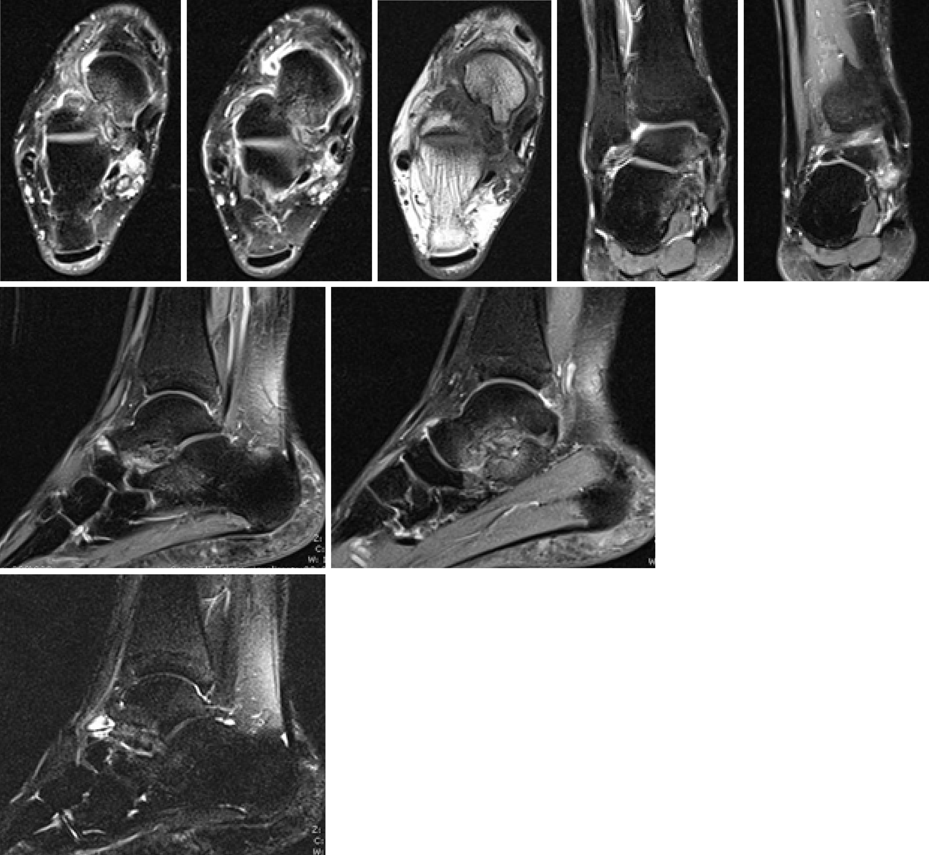
Figure 5 Magnetic resonance imaging of the ankle of Patient 2.
Loose bodies and degenerative arthritis are shown.
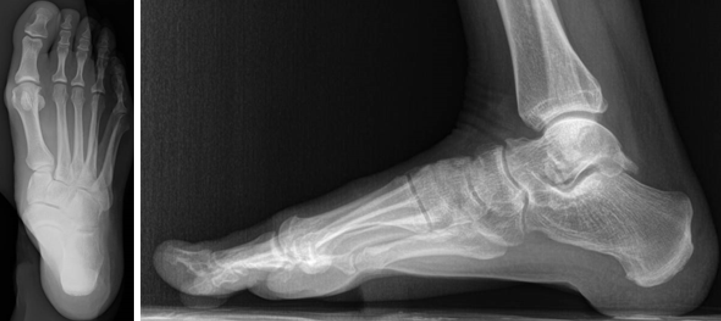
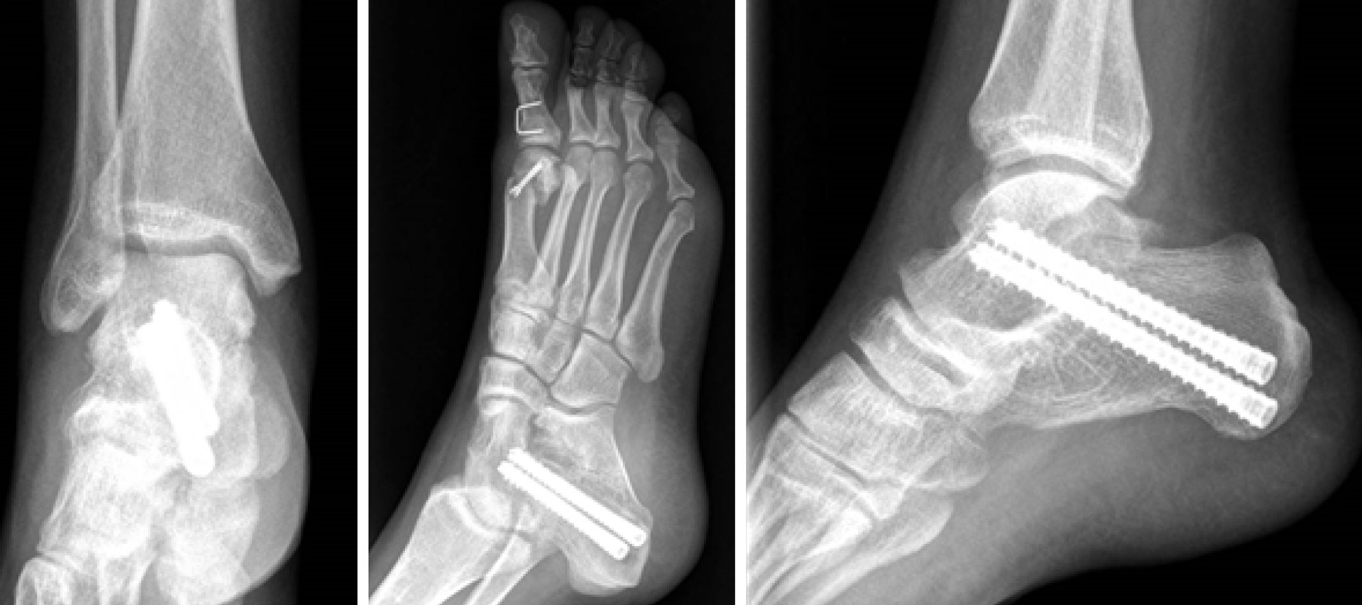
Figure 6 Post-operative radiographs of Patient 2: Synovectomy, removal of loose bodies and arthrodesis of subtalar joint is performed.
Youngswick and Akin osteotomies are performed to correct hallux valgus.
- Citation: Monestier L, Riva G, Stissi P, Latiff M, Surace MF. Synovial chondromatosis of the foot: Two case reports and literature review. World J Orthop 2019; 10(11): 404-415
- URL: https://www.wjgnet.com/2218-5836/full/v10/i11/404.htm
- DOI: https://dx.doi.org/10.5312/wjo.v10.i11.404


