Copyright
©The Author(s) 2019.
World J Orthop. Oct 28, 2019; 10(11): 394-403
Published online Oct 28, 2019. doi: 10.5312/wjo.v10.i11.394
Published online Oct 28, 2019. doi: 10.5312/wjo.v10.i11.394
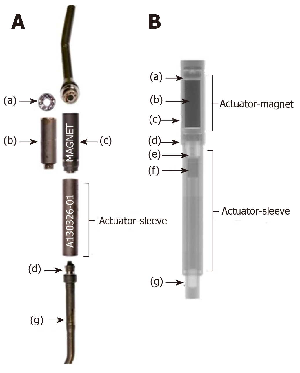
Figure 1 Clinical and radiographic image of a magnetically controlled growing rod after sectioning.
A: Clinical image of a magnetically controlled growing rod after sectioning; B: Radiographic image of a magnetically controlled growing rod after sectioning. The keeper plate (label c) is seen in its position around the magnet (label b). The Figure is adapted from Panagiotopoulou et al[31].
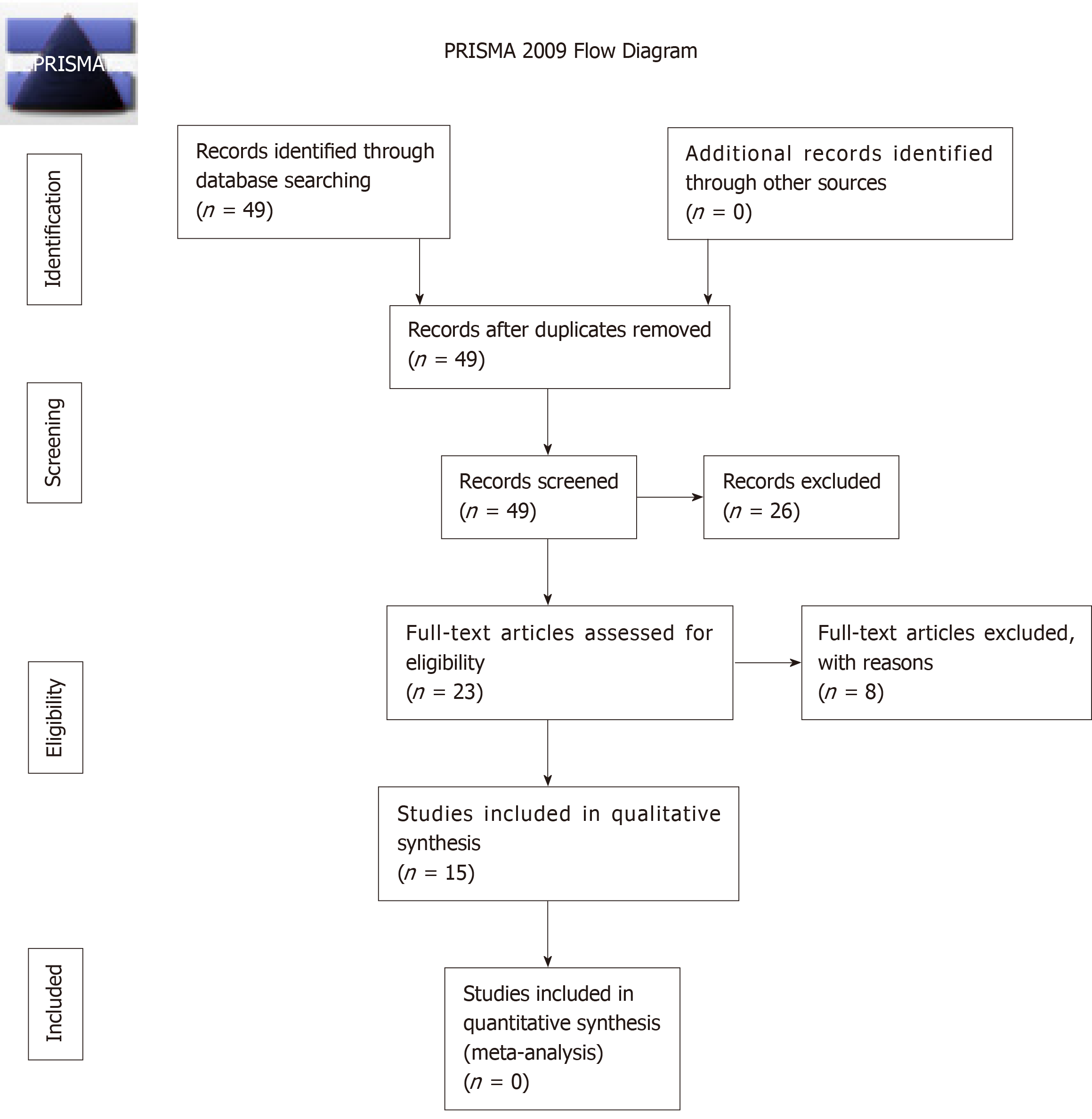
Figure 2 The preferred reporting items for systematic reviews and meta-analyses flowchart depicting protocol for reviewing studies considered for inclusion.
PRISMA: Preferred reporting items for systematic reviews and meta-analyses.
- Citation: Shaw KA, Hire JM, Kim S, Devito DP, Schmitz ML, Murphy JS. Magnetically controlled growing instrumentation for early onset scoliosis: Caution needed when interpreting the literature. World J Orthop 2019; 10(11): 394-403
- URL: https://www.wjgnet.com/2218-5836/full/v10/i11/394.htm
- DOI: https://dx.doi.org/10.5312/wjo.v10.i11.394


