Copyright
©The Author(s) 2019.
World J Orthop. Jan 18, 2019; 10(1): 33-44
Published online Jan 18, 2019. doi: 10.5312/wjo.v10.i1.33
Published online Jan 18, 2019. doi: 10.5312/wjo.v10.i1.33
Figure 1 Case timeline.
CMS: Constant-Murley score.
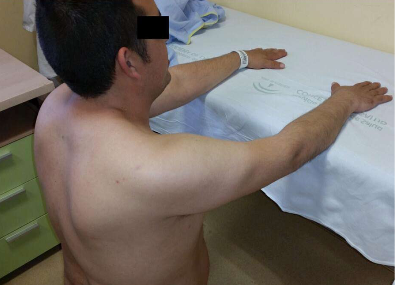
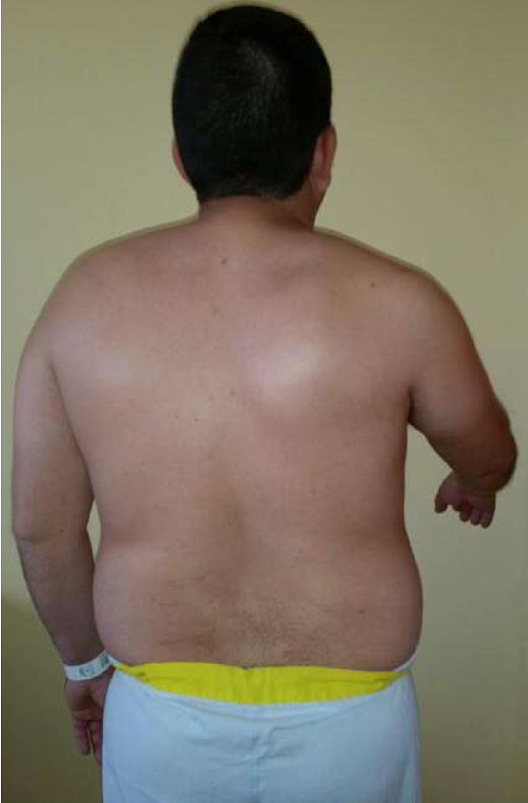
Figure 2 Scapular winging with right shoulder in passive flexion.
Figure 3 Decreased shoulder movement with 60º active flexion.
Scapular winging is observed with shoulder flexion.
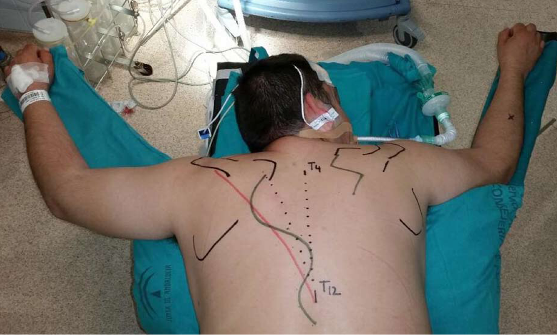
Figure 4 Superficial anatomical landmarks and incision planning.
Figure 5 Bone tissues can be seen at the tips of the mosquito clamp coming from T11 and T12 spinal processes.
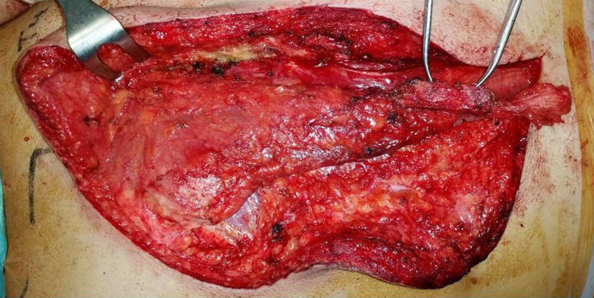
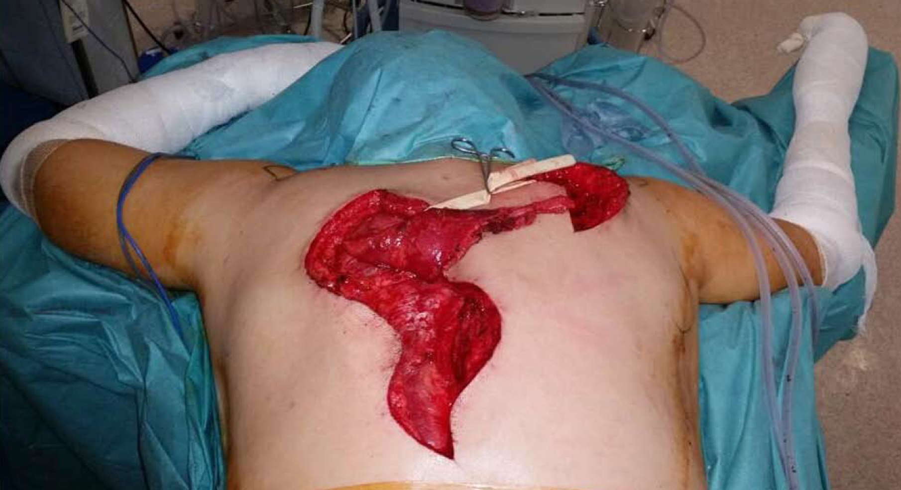
Figure 6 The compound osteomuscular trapezius flap was raised and passed through a subcutaneous tunnel (Penrose drain) to the second incision.
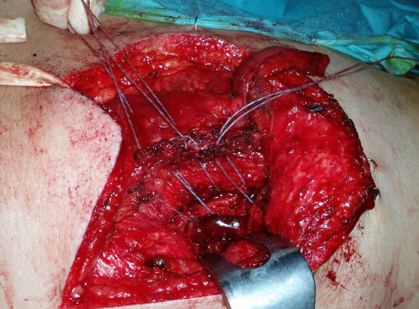
Figure 7 The flap was fixed using anchors in the beds and transosseous sutures through the scapular spine to attach the tendon to the footprint.
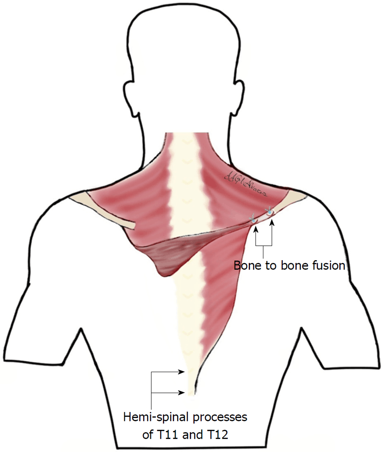
Figure 8 Diagram of the contralateral trapezius compound osteomuscular flap transfer.
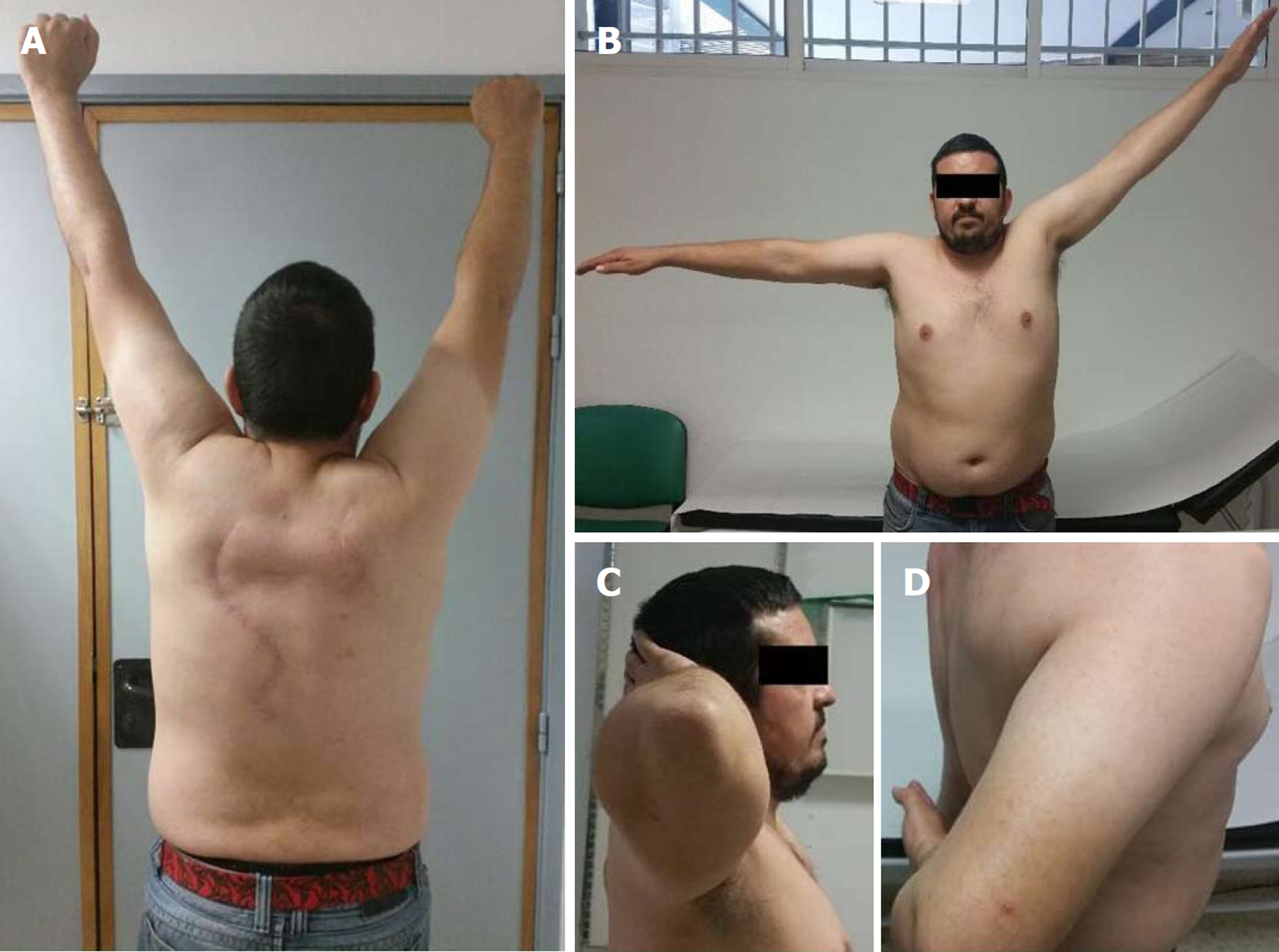
Figure 9 Active range of motion at the 6-mo follow-up.
A: Flexion (notice flap working); B: Abduction; C: External rotation; D: Internal rotation and normal lift-off test.
- Citation: Gil-Álvarez JJ, García-Parra P, Anaya-Rojas M, Martínez-Fuentes MDP. Contralateral trapezius transfer to treat scapular winging: A case report and review of literature. World J Orthop 2019; 10(1): 33-44
- URL: https://www.wjgnet.com/2218-5836/full/v10/i1/33.htm
- DOI: https://dx.doi.org/10.5312/wjo.v10.i1.33


