INTRODUCTION
Induction of apoptosis by chemotherapeutics is considered as one of the major anti-cancer effects leading to inhibition of tumour growth. Over the past years our understanding of signaling pathways associated with induction of apoptosis, and knowledge on executioners of apoptosis, has substantially increased. Recently, cell-to-cell stochastic variability has become central to apoptotic cell death signaling[1-6]. Computational models are well suited to provide mechanistic insight into the system level regulations of apoptosis signaling and its large cellular variability. Studies that are possible using in silico approaches might be inaccessible by other techniques. Recent novel findings in the area of apoptotic cell death signaling can have far-reaching implications in cancer biology and therapy. Our computational efforts, in synergy with parallel biological experiments, attempt to explore some of the fundamental issues in cancer biology within this new paradigm of apoptosis signaling.
DEVELOPING COMPUTATIONAL MODELS OF CELL DEATH SIGNALING FOR NORMAL AND CANCER CELLS
Monte Carlo models are generally suitable for simulating inherent stochasticity of signaling reactions in complex signaling pathways[1,7]. Our recent work has elucidated that cell-to-cell stochastic variability is a fundamental characteristic of apoptosis signaling and a significant part of such variability can arise due to inherent stochastic fluctuations of chemical reactions[1,6,8,9]. Even when the intrinsic stochastic variability is not dominant, Monte Carlo models have the advantage over ordinary differential equation (ODE) based models, as they can simulate spatial heterogeneity in an explicit manner. Examples of such spatial localizations in apoptosis signaling include translocation of activated Bax molecules onto mitochondrial outer membrane, release of cytochrome c from mitochondria to cytosol and redistribution of Apaf-1 in the cytosol, all of which depend on the cell type and the level of Bcl-2 proteins[10,11]. In addition, we could easily incorporate realistic variations in (1) protein concentrations that may arise from stochastic gene regulations[3,12-15]; and (2) reaction rate constants, for example, due to variations in pH in the cytosolic environment. In our initial studies we grouped functionally redundant proteins so that a single representative protein simulates all proteins of similar function that are possibly expressed within a given cell type. For example, apoptotic inhibitor Bcl-2 captured the effect of all the inhibitory Bcl-2 like proteins[1,9]. In the future we plan to simulate a more cell-type-specific signaling network of apoptosis, the results of which can be readily compared with data obtained from parallel biological experiments for specific cell types. Such an expanded signaling network will often involve signaling species with low concentrations at initial time, or dynamically generate a few molecules due to specific inhibitory reactions, leading to inherent stochastic fluctuations that can be best studied using stochastic approaches.
DIFFERENTIAL SIGNALING THROUGH EXTRINSIC AND INTRINSIC PATHWAYS OF APOPTOSIS
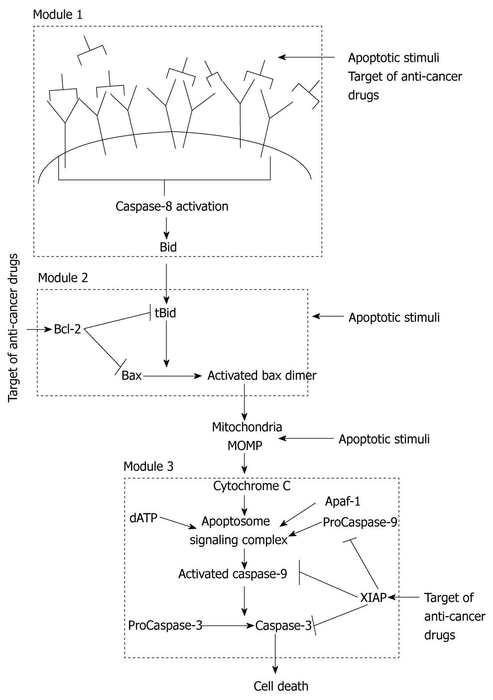
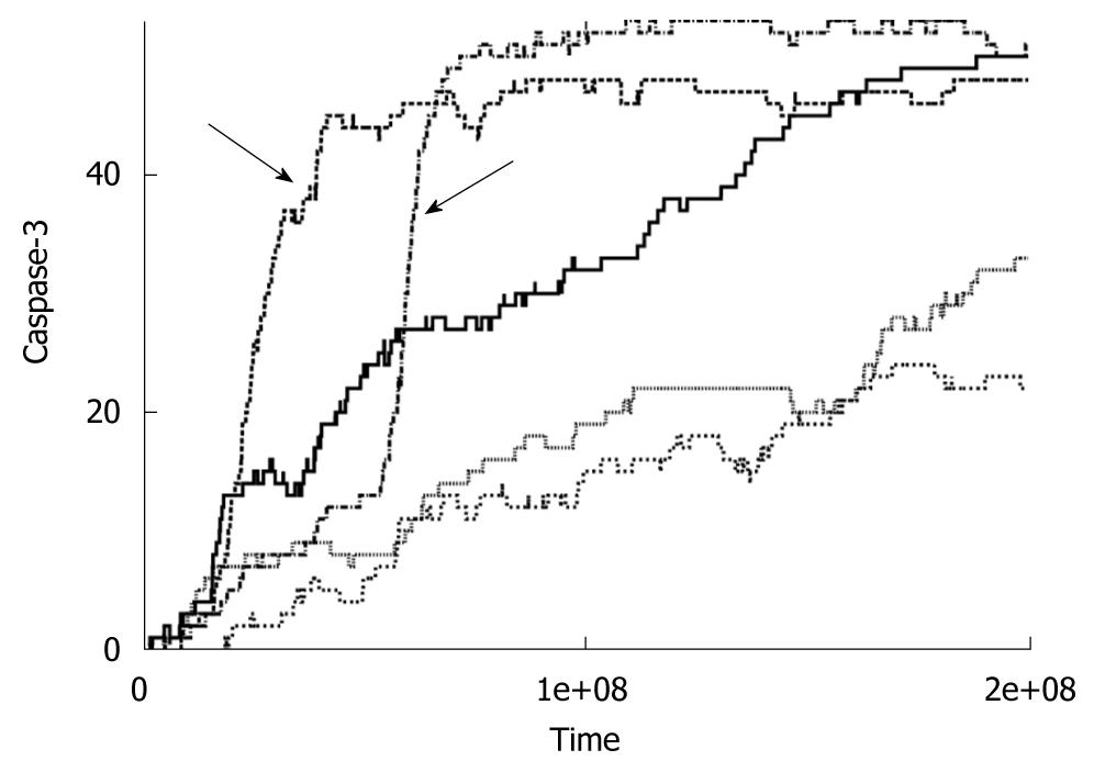
Apoptosis is regulated through two distinct signaling pathways that are joined in a global loop structure as both pathways converge on activation of effector Caspase-3[10]. The extrinsic (also called the type 1) pathway directly activates Caspase-3 by enzymatic reactions catalyzed by activated Caspase-8 molecules. The intrinsic (also called the type 2) pathway is regulated by mitochondrial cytochrome c release and apoptosome formation. We can assume that three local signaling modules coordinate apoptosis in the type 2 pathway (Figure 1). Cell death can be induced by an apoptotic stimulus acting at any of the three different signaling modules (or right before them) of the apoptotic pathway (Figure 1): (1) death ligand binding and Caspase-8 activation; (2) Bax activation by BH3 only proteins; and (3) Ca2+ release that activates only the post-mitochondrial events. Our initial studies showed that, for the case of Caspase-8-mediated apoptosis, concentration of active Caspase-8 decides between the two pathways of apoptosis. Membrane reorganization, such as clustering of death receptors in raft signaling domains, determines the level of Caspase-8 activation in a cell type specific manner[16]. For large concentrations of Caspase-8, direct Caspase-3 activation occurs in a fast (minutes) deterministic manner. Such rapid activation of apoptosis has been observed in various cell types due to Fas ligand binding to Fas receptor[17]. Decrease in the strength of an apoptotic stimulus begins to activate the intrinsic pathway (Figure 2), as the rate constant for Caspase-8-Bid interaction is higher than that for Caspase-8-Capsase 3 association[1,18].
Figure 1 Schematic representation of the apoptosis signaling pathway that indicates existence of three distinct signaling modules in the apoptotic intrinsic pathway.
Apoptosis can be activated at various locations in the intrinsic pathway. We also show some of the targets of cancer drugs.
Figure 2 Time course of caspase-3 activation for low concentrations of caspase-8 (< 5 nmol).
Data is shown for five individual cells. Arrows indicates switch to the intrinsic pathway of apoptosis at the level of single cells.
LARGE CELL-TO-CELL VARIABILITY THROUGH THE INTRINSIC (MITOCHONDRIAL) PATHWAY CAN EXPLAIN SLOW APOPTOSIS
We observe slow apoptosis (hours) when low concentrations of Caspase-8 are used in our simulations. We also find large cell-to-cell stochastic variability in the case of slow apoptosis. Similar signaling behavior is observed, irrespective of the Caspase-8 concentration used, when we set the kinetic constants for the type 1 pathway to zero, confirming that slow apoptosis is a characteristic of the intrinsic pathway of apoptosis signaling. Such a study of pure type 2 apoptosis can be carried out in silico in a clean manner but will be difficult to achieve in biological experiments. When we perturbed the intrinsic apoptotic pathway downstream of Caspase-8 activation, we kept observing slow apoptosis with large cell-to-cell variability. Our results seem to explain very slow (1-100 h) apoptosis observed experimentally under a variety of conditions, for various types of cells and apoptotic stimuli, including under oxidative stress[2-6,9,10,19,20]. Additional variations in protein concentrations in our simulations act in tandem with intrinsic stochasticity of signaling reactions to enhance cell-to-cell variability in apoptosis. Caspase-3 activation occurs in an all-or-none (digital) manner for single cells implicating signaling amplification of a weak stimulus through the intrinsic pathway. However, the information of strength of the stimulus is contained in the time-to-death and its cell-to-cell variability. Large cell-to-cell variability with all-or-none type Caspase-3 activation, as observed in our simulations, resulted in bimodal probability distributions for Caspase-3 activation that are thought to be characteristic of apoptosis signaling through the intrinsic pathway[1]. Later experiments confirmed existence of such bimodal probability distributions in Caspase-3 activation[2,3,6,9].
MINIMAL MODEL OF A SIGNALING NETWORK DEMONSTRATES CELL-TO-CELL VARIABILITY IN APOPTOSIS IN A CELL-TYPE INDEPENDENT MANNER
In parallel, we derived a minimal model of a signaling network that is designed to sense an external stimulus and respond to it in an adaptive manner[8]. This minimal network is derived based on some simple assumptions on its signaling response without any prior knowledge of the apoptotic pathway. A three-step fast-slow-fast pathway in the minimal network was shown to be sufficient to generate large cell-to-cell variability as observed in our Monte Carlo simulations of the intrinsic pathway of apoptosis[1]. This minimal network also captures the change from rapid deterministic to slow stochastic signaling as the strength of the stimuli is varied, and a quantitative estimation of the threshold stimulus is obtained. This could be potentially important if one wants to engineer cancer cells to convert from type 2 to type 1 for fast apoptotic activation. The crucial slow reaction in the intermediate step of the minimal network can mimic the slow activation of Bax or the apoptosome formation in apoptosis signaling. Thus, we can infer that some of the pertinent qualitative features of apoptosis signaling, as observed in our Monte Carlo simulations, are cell type independent. Such a conclusion is significant given the fact that cellular protein levels and even the type of molecules present in the apoptotic pathways vary significantly among cell types[3,10,12]. Cancer cells are known to over-express a variety of apoptotic inhibitors, which confer them unusual resistance to apoptosis[21-24]. The level of over-expression varies significantly among cancer sub-types and even among patients having similar sub-types[25,26].
HOW APOPTOTIC INHIBITORS PROVIDE PROTECTION TO CANCER CELLS: IMPLICATIONS FOR CANCER THERAPY
In a recent study, we have shown that over-expression of Bcl-2 like proteins can slow down apoptosis and increase cell-to-cell stochastic variability[9]. A high Bcl-2 level allows activation of only a few Bax molecules under apoptotic stimuli and thus dynamically generates mechanisms for stochastic fluctuations caused by small number of molecules. Interestingly, cancer cells are often primed for death by increasing the levels of apoptotic BH3 proteins. However, in such cells, apoptosis is kept in check by continuous inhibition by anti-apoptotic Bcl-2-like proteins[22-24]. Bcl-2 binds with several pro-apoptotic molecules creating a local loop structure (signaling module 2) in the intrinsic pathway that leads to non-linear and stochastic effects in its inhibitory action. Our simulations demonstrate that, beyond a threshold level, Bcl-2 imparts a strong inhibitory effect on apoptosis and thus can explain apoptosis resistance of cancer cells. For normal cells, having over-expressed Bcl-2 proteins, prolonged time-to-death might provide an opportunity for a particularly slow cell to acquire tumor initiating features. Behavior similar to tBid-Bcl-2-Bax signaling (in module 2) might be observed downstream of mitochondria (in signaling module 3) where higher Apaf-1 level might make cancer cells prone to apoptotic death, although such an effect can be abolished by the dominant effect of increased X-linked inhibitor of apoptosis protein (XIAP) levels[27]. XIAP also has multiple binding partners in a local loop network structure and contributes to generation of highly non-linear and stochastic signaling. Hence, pre- and post- mitochondrial events in the intrinsic pathway are heavily regulated by two different loop network structures in two distinct signaling modules (Figure 1). Computational models are well-suited to elucidate mechanisms of non-linear and stochastic signaling through those signaling modules. As a result, such models can help design optimal strategies to perturb those signaling modules by making use of the inherent apoptotic vulnerability of cancer cells. Initial simulations show increased cell death only for cancer cells over-expressing BH3 protein Bid (unpublished observations), under a single agent treatment scenario, such as under the action of Bcl-2 inhibitor HA14-1[28-30]. Our computational studies can clarify the basis of such inherent vulnerability of cancer cells for all three signaling modules (Figure 1). However, targeting only a single module (Figure 1), for example ligation of death receptors at the signaling module 1, will provide an opportunity for a significant number of cells to escape death. Such fractional killing of cancer cells occurs not only due to cellular variations in protein levels but also from inherent stochastic variability in signaling reactions[9]. Computational modeling was well-suited to establish that inherent stochastic variability by itself, even when all the other cellular parameters remain identical, can generate large cell-to-cell variability, comparable to that observed in apoptosis activation experiments[9]. Such large cell-to-cell variability in time-to-death provides an opportunity for opposing growth signals to up-regulate downstream apoptotic inhibitors such as XIAP. This is particularly relevant as apoptosis activation under chemotherapeutic treatment can be slow enough to allow synthesis of inhibitor proteins of varying concentrations through stochastic gene regulations. Targeting multiple signaling modules simultaneously, using combined treatments, can be effective in reducing stochastic effects and fractional killing of cancer cells. Computational studies can provide us with a range of concentrations for optimal induction of apoptosis in a combined treatment scenario and can guide the design of biological experiments. We are currently exploring the combined effect of HA14-1, an inhibitor of Bcl-2[28-30], and embelin[27], an inhibitor of XIAP, that can induce apoptotic collapse in cancer cells.
Peer reviewers: George G Chen, MD, PhD, Professor, Department of Surgery, The Chinese University of Hong Kong, Prince of Wales Hospital, Shatin, Hong Kong, China; Guido Cavaletti, MD, Associate Professor, Department Neuroscienze e Tecnologie Biomediche - Università di Milano “Bicocca”, v. Cadore 48 - 20052 Monza, Italy
S- Editor Cheng JX L- Editor Negro F E- Editor Ma WH


