Topic Highlight Open Access
Copyright ©2010 Baishideng Publishing Group Co., Limited. All rights reserved.
World J Clin Oncol. Nov 10, 2010; 1(1): 12-17
Published online Nov 10, 2010. doi: 10.5306/wjco.v1.i1.12
Molecular mechanism of base pairing infidelity during DNA duplication upon one-electron oxidation
Jóhannes Reynisson, Department of Chemistry and Auckland Bioengineering Institute, The University of Auckland, Auckland 1142, New Zealand
Author contributions: Reynisson J solely contributed to this paper.
Correspondence to: Dr. Jóhannes Reynisson, Department of Chemistry and Auckland Bioengineering Institute, The University of Auckland, Private Bag 92019, Auckland 1142, New Zealand. j.reynisson@auckland.ac.nz
Telephone: +64-9-3737599 Fax: +64-9-3737422
Received: April 16, 2010
Revised: September 21, 2010
Accepted: September 28, 2010
Published online: November 10, 2010

Abstract

The guanine radical cation (G•+) is formed by one-electron oxidation from its parent guanine (G). G•+ is rapidly deprotonated in the aqueous phase resulting in the formation of the neutral guanine radical [G(-H)]. The loss of proton occurs at the N1 nitrogen, which is involved in the classical Watson-Crick base pairing with cytosine (C). Employing the density functional theory (DFT), it has been observed that a new shifted base pairing configuration is formed between G(-H) and C constituting only two hydrogen bonds after deprotonation occurs. Using the DFT method, G(-H) was paired with thymine (T), adenine (A) and G revealing substantial binding energies comparable to those of classical G-C and A-T base pairs. Hence, G(-H) does not display any particular specificity for C compared to the other bases. Taking into account the long lifetime of the G(-H) radical in the DNA helix (5 s) and the rapid duplication rate of DNA during mitosis/meiosis (5-500 bases per s), G(-H) can pair promiscuously leading to errors in the duplication process. This scenario constitutes a new mechanism which explains how one-electron oxidation of the DNA double helix can lead to mutations.

Key Words: Base pairing; Density functional theory; Deprotonation; DNA duplication; Duplication rate; Guanine neutral radical; Nucleotides; Oxidative DNA damage; Radical lifetime


INTRODUCTION

The aqueous redox chemistry of the nucleosides and nucleotides has been extensively investigated for the last 40 years using pulse radiolysis, laser photolysis, electron spin resonance and other time resolved and steady state techniques[1]. More recently, theoretical methods have been employed in the study of redox damage of DNA[1]. This intense interest in the components of DNA is understandable since it carries our genetic code and if damaged can lead to mutations possibly resulting in cancer[2,3]. Furthermore, oxidative damage of DNA is implicated in aging[4] and bacterial drug resistance[5]. It is now understood that DNA damage initiated by ionising radiation elicits a complicated set of events engaging various signalling pathways in cells[6].

Given that cumulative cancer risk increases with the fourth power of age and is associated with an accumulation of DNA damage, oxidative DNA damage is of great interest regarding early tumorigenesis and eventually cancer. These redox damage mechanisms have a potential role in the initiation, promotion and malignant conversion stages of carcinogenesis[2]. Lesions such as 7,8-dihydro-8-oxoguanine (8OG) are established biomarkers of oxidative stress; coupled with their mutagenicity in mammalian cells, this has led to them being proposed as intermediate markers of cancer[2]. A more complete understanding of these oxidative damage processes in DNA is highly desirable in order to find new therapeutic strategies to battle this devastating disease.

BINDING SPECIFICITY ALTERATION OF THE GUANINE BASE

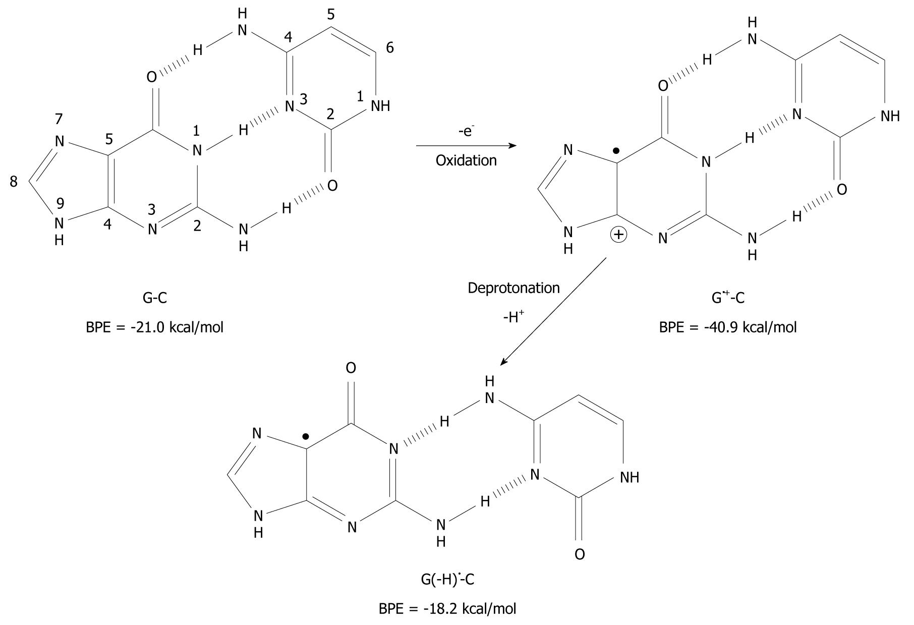
It has been found that when organic molecules are one-electron oxidized in the aqueous phase, a rapid deprotonation occurs from hydrogen bond donors undoubtedly driven by the massive solvation energy of the proton (∆Gaq = -263.9 kcal/mol)[7-9]. As an example, the pKa-value of cytosine (C) is lowered from 12.15 to between 2 and 4 when C is one-electron oxidized[8,10,11]. With respect to DNA, guanine (G) is its most easily oxidized component[12] and when the π-stack of double stranded DNA loses an electron, the positive charge migrates to G-C rich areas in the double strand[13-16] and the pKa-value of G is lowered significantly from 9.4 to 3.9 at the nitrogen-1 atom (N1), as depicted in Figure 1[10,17,18]. After departure of the proton from the N1-site, it becomes a hydrogen bond acceptor instead of a hydrogen bond donor. The question has emerged as to whether this event leads to a change in the pairing ability of the G moiety with other bases[19]? In fact, it is a common view that ligand hydrophobicity improves affinity, whereas hydrogen bonding improves specificity for interactions in biochemical systems[20]. Simulating one-electron oxidation and the consequent deprotonation of the central N1-proton for G-C, using the density functional theory (DFT)[21], a new slipped conformation of the base pair was formed as depicted in Figure 2[19]. This slipped configuration, G(-H)-C, was later independently derived by Bera et al[22] using a systematic search for all possible hydrogen bonding configurations between G(-H) and C. The predicted base pairing energy (BPE) was -18.2 kcal/mol for G(-H)-C[19,23]. This lies between the BPE’s of the adenine-thymine base pair (A-T) at -13.0 kcal/mol and that of G-C at -21.0 kcal/mol[24,25].

Figure 1
Figure 1 Oxidation of guanine (G) and deprotonation of its radical cation (G•+). The pKa-value of G is drastically lowered upon one-electron oxidation and subsequent deprotonation of the N1 proton changes it from a hydrogen bond donor to a hydrogen bond acceptor. The number of atoms constituting G is shown. Drib: 2’-deoxyribose moiety.
Figure 2
Figure 2 Deprotonation-induced structural change of the G-C base pair initiated by one-electron oxidation leading to the shifted base pair G(-H) - C. BPE: Base pairing energy.
DEPROTONATION OF OXIDIZED GUANINE IN DOUBLE STRANDED DNA

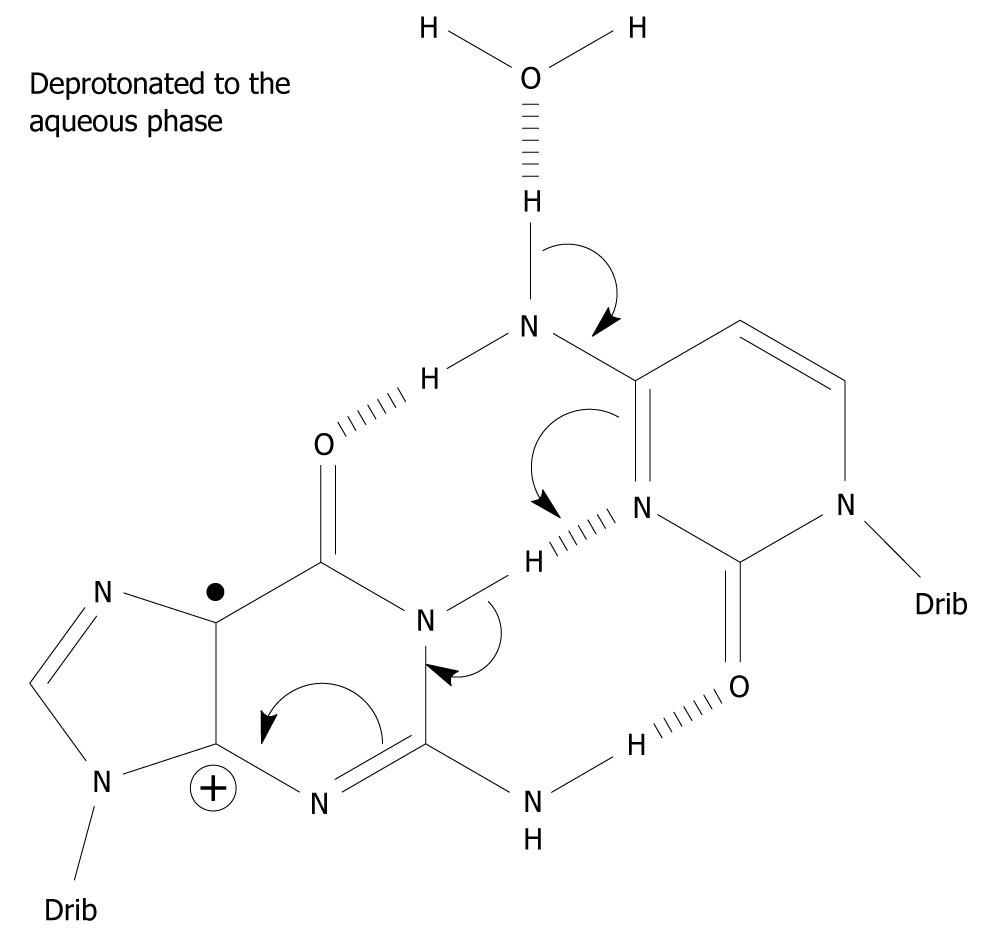
Under what circumstances can G•+-C in the DNA stack lose the central N1 proton making up one of the Watson-Crick hydrogen bonds? It does not have access to the aqueous phase since it is the central hydrogen bond and is flanked by base pairs on either side in the double stranded DNA helix. It is imperative that N1-H comes into contact with the water phase (water acting as a proton acceptor), i.e. within G-C, the G(N1-H)-C(N3) Watson-Crick hydrogen bond has to be broken for the N1 proton to be lost (Figure 2). The hydrogen bonds between the base pairs may be broken in three situations: First, the “swing-out” of the bases by concerted thermal motions of the DNA strand[26,27]. This mechanism is unlikely since it takes place on the milli- to micro-second time scale and is in competition with further charge migration in the DNA helix and/or with water addition to C8 of G•+, which are considerably faster. The rate of charge migration is estimated as 5 × 10⁷/s and 6 × 10⁴/s for the water addition, i.e. in the micro-nanosecond timescale[16,28]. Furthermore, the BPE of G•+-C is increased to -40.9 kcal/mol compared to -21.0 kcal/mol of its parent pair, inhibiting the frequency of the breathing motions of the base pair[24,29,30]. Second, when duplication of DNA occurs, the DNA strand is untwisted and the hydrogen bonds between the bases are broken to allow duplication of the strand. Third, during DNA transcription to messenger-RNA, it proceeds in a similar fashion to the duplication of DNA. In addition, it has been suggested that deprotonation occurs from the exocyclic amine group of C in G•+-C based on pulse-radiolysis and kinetic isotope experiments[31-33]. The proposed deprotonation mechanism is shown in Figure 3. This reaction cascade can lead to the G(-H)-C slipped configuration[34].

Figure 3
Figure 3 A possible mechanism which involves the exocyclic amine moiety on C as the proton donor of the one-electron oxidized base pair in which the initial charge sits on G, i. e. in the complementary strand. Spin-charge separation between G and C plays a crucial role in the reaction cascade. The depicted deprotonation can lead to the formation of G(-H)-C[34].
PAIRING INFIDELITY OF THE DEPROTONATED GUANINE RADICAL

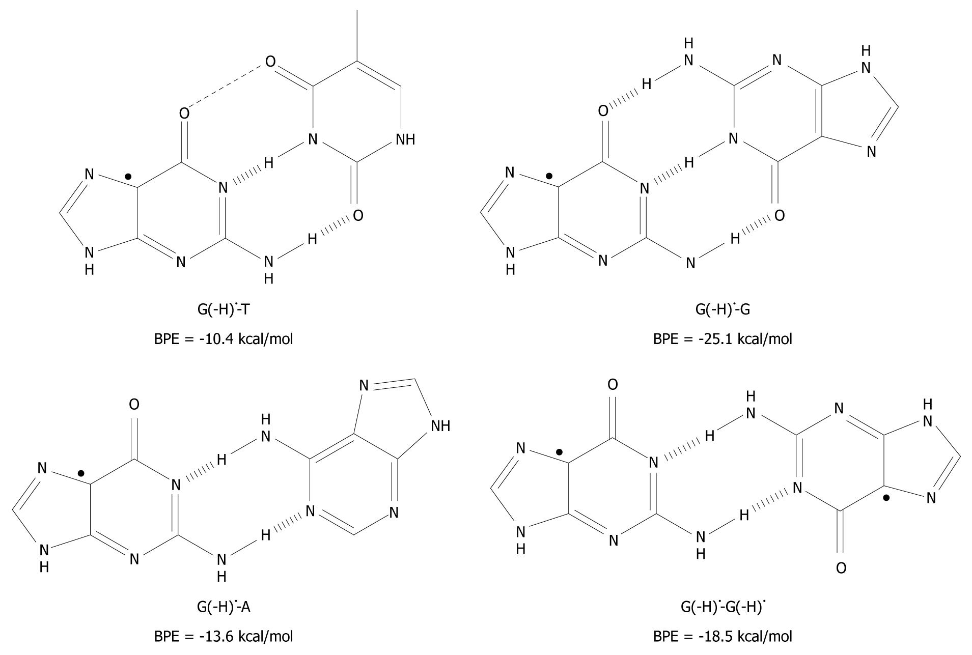
A related question has emerged as to whether it is possible to pair T, A and G itself to G(-H) ? This was investigated using the DFT method and the results are given in Figure 4[19].

Figure 4
Figure 4 The unnatural base pairs between G(-H) and the other bases[19]. The substantial base pairing energy (BPE) for the non-classical complexes depicted leads to the conclusion that G(-H) does not have any specificity for C.

Armed with the knowledge that the G(-H)-C base pair has only two hydrogen bonds, G(-H) was paired to T and structurally optimized. The BPE was calculated to be -10.4 kcal/mol for G(-H)-T, which is comparable to the A-T base pairing energy (-13.0 kcal/mol[24,25,29,35]). The relatively low energy can be explained in terms of the non-planarity of the bases with respect to each other. On the basis of the calculations, they are roughly 25º out of plane, measured at their carbonyl groups, O6 (G) and O4 (T). The distance between these oxygen atoms is 3.5 Å, which proximity leads to Coulombic repulsion and hence the non-planar conformation.

The calculated hydrogen bonding energy of the G(-H)-A base pair is -13.6 kcal/mol, as shown in Figure 4. This binding is somewhat stronger than that for the natural A-T pairing (-13.0 kcal/mol)[24,25,29,35].

The hydrogen bond energy of G(-H)-G (structure depicted in Figure 4) is similar to that of G-C[36]. This is not surprising as three hydrogen bonds are formed in both structures. A second type of G-G base pair is conceivable between two G(-H) moieties (G(-H)-G(-H)) as shown in Figure 4. For this, the hydrogen bond energy is -18.5 kcal/mol, somewhat lower than for G(-H)-G, since it has one less hydrogen bond. The Pt(II) electrophile coordinates at N7 of G. This acidifies the N1 proton, similar to the oxidation of G. With these Pt-G species, structures similar to G(-H)-G and G(-H)-G(-H) were observed with 1H-NMR and X-ray crystallography[37], which provides experimental evidence of their existence.

ONE-ELECTRON OXIDATION DURING DNA DUPLICATION

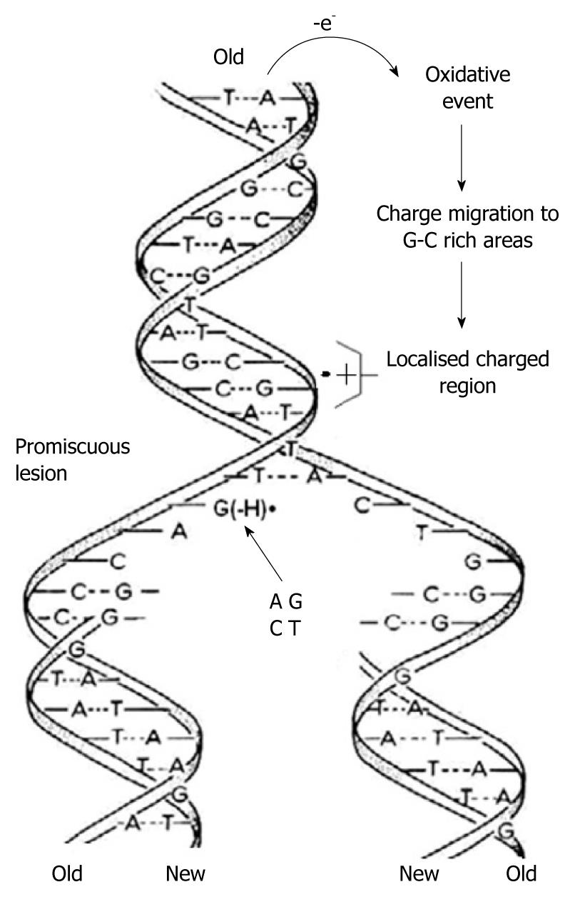
Using in-situ photolysis electron paramagnetic resonance (EPR), Hildenbrand and Schulte-Frohlinde, detected a long-lived radical (lifetime 5 s) which was produced only from double stranded DNA when ionised with < 220 nm light in an aqueous solution at pH 7[38]. This radical was assigned to G(-H). The rate of DNA duplication was measured to be between 5-500 nucleotides/s depending on the cell type, species and other factors[39,40]. Considering the long lifetime of G(-H) in double stranded DNA and the rapid DNA duplication rate, it emerges that in the case of one-electron oxidation during mitosis-/-meiosis, G(-H) is formed when the two strands unwind. As shown in Figure 4, it can form base pairs with all of the nucleotides with binding energies similar to the classical A-T and G-C Watson-Crick base pairs. This means that G(-H) does not have specific affinity for C, i.e. it is completely promiscuous when it comes to base pairing. Therefore G(-H) can pair with all of the nucleotides leading to mispairing. A depiction of this scenario is presented graphically in Figure 5.

Figure 5
Figure 5 As the two strands of the double helix unwind, each pairs up with the appropriate bases to form a new double helix. The two new helices are identical to each other and to the original. This process is compromised by one-electron oxidation of the π-DNA stack, deprotonation from G•+ and the subsequent formation of G(-H), which is promiscuous with regard to base pairing.

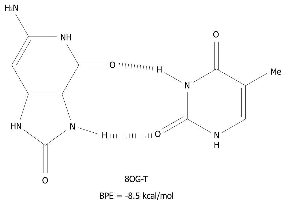
The mechanism presented here is new and an alternative to the scenario that mispairing of DNA bases is mostly caused by oxidative end products such as 8OG[41]. These products are closed shell, i.e. they are not radical species and therefore, have a much longer lifetime than G(-H). 8OG is one of the many redox products which is derived from the oxidation, and the subsequent water addition, of G[17,18,42]. It can form syn-anti base pairs[43], with all of the nucleotides and these have base pairing energies of -10 kcal/mol[44]. The 8OG-T base pair is depicted in Figure 6 as an example of syn-anti base pairs.

Figure 6
Figure 6 The syn-anti base pair of 8-oxoguanine-T. BPE: Base pairing energy; 8OG: 8-oxoguanine.

So far, the role of DNA polymerase has not been considered and the DNA bases and base pairs have been treated as in vacuo as a model. The structure of DNA polymerase and its steric limitations within the active site are well documented[45-47]. The structure of the binding site in the replicating enzymes will undoubtedly have an effect on the proposed infidelity mechanism based on G(-H), e.g. the rate of duplication.

CONCLUSION

In this review, an alternative mechanism for promiscuous base pairing during DNA duplication, initiated by one-electron oxidation, is proposed based on theoretical calculations. Some experimental results exist which support the existence of the non-classical base pairs discussed, i.e. the slipped G(-H)-C and the G(-H)-G base pairs. Further experimental and theoretical work is needed to corroborate the mechanism proposed. In particular, experiments conducted with time resolved resonance Raman spectroscopy on model DNA duplication systems are pertinent as well as modelling studies on the effect of DNA polymerase.

Footnotes

Peer reviewer: Millie M Georgiadis, Associate Professor, Department of Biochemistry and Molecular Biology Indiana University School of Medicine, 635 Barnhill Dr. Indianapolis, IN 46202, United States

S- Editor Cheng JX L- Editor Webster JR E- Editor Ma WH

References
1.  von Sonntag C Free-Radical-Induced DNA Damage and Its Repair. A Chemical Perspective. Springer-Verlag: Berlin Heidelberg 2006; .  [PubMed]  [DOI]  [Cited in This Article: ]
2.  Cooke MS, Evans MD, Dizdaroglu M, Lunec J. Oxidative DNA damage: mechanisms, mutation, and disease. FASEB J. 2003;17:1195-1214.  [PubMed]  [DOI]  [Cited in This Article: ]
3.  Arlt VM. 3-Nitrobenzanthrone, a potential human cancer hazard in diesel exhaust and urban air pollution: a review of the evidence. Mutagenesis. 2005;20:399-410.  [PubMed]  [DOI]  [Cited in This Article: ]
4.  Merry BJ. Oxidative stress and mitochondrial function with aging-the effects of calorie restriction. Aging Cell. 2004;3:7-12.  [PubMed]  [DOI]  [Cited in This Article: ]
5.  Kohanski MA, DePristo MA, Collins JJ. Sublethal antibiotic treatment leads to multidrug resistance via radical-induced mutagenesis. Mol Cell. 2010;37:311-320.  [PubMed]  [DOI]  [Cited in This Article: ]
6.  Darzynkiewicz Z, Traganos F, Wlodkowic D. Impaired DNA damage response-an Achilles’ heel sensitizing cancer to chemotherapy and radiotherapy. Eur J Pharmacol. 2009;625:143-150.  [PubMed]  [DOI]  [Cited in This Article: ]
7.  Steenken S. Purine bases, nucleosides, and nucleotides: aqueous solution redox chemistry and transformation reactions of their radical cations and e- and OH adducts. Chem Rev. 1989;89:503-520.  [PubMed]  [DOI]  [Cited in This Article: ]
8.  Steenken S. Electron-transfer-induced acidity/basicity and reactivity changes of purine and pyrimidine bases. Consequences of redox processes for DNA base pairs. Free Radic Res Commun. 1992;16:349-379.  [PubMed]  [DOI]  [Cited in This Article: ]
9.  Tissandier MD, Cowen KA, Feng WY, Gundlach E, Cohen MH, Earhart AD, Coe JV. The proton's absolute aqueous enthalpy and gibbs free energy of solvation from cluster-ion solvation data. J Phys Chem A. 1998;102:7787-7794.  [PubMed]  [DOI]  [Cited in This Article: ]
10.  Dean JA Lange‘s Handbook of Chemistry. New York: McGraw-Hill 1985; .  [PubMed]  [DOI]  [Cited in This Article: ]
11.  Geimer J, Hildenbrand K, Naumov S, Beckert D. Radicals formed by electron transfer from cytosine and 1-methylcytosine to the triplet state of anthraquinone-2,6-disulfonic acid. A Fourier-transform EPR study. Phys Chem Chem Phys. 2000;2:4199-4206.  [PubMed]  [DOI]  [Cited in This Article: ]
12.  Steenken S, Jovanovic SV. How Easily Oxidizable Is DNA? One-Electron Reduction Potentials of Adenosine and Guanosine Radicals in Aqueous Solution. J Am Chem Soc. 1997;119:617-618.  [PubMed]  [DOI]  [Cited in This Article: ]
13.  Steenken S. Electron transfer in DNA? Competition by ultra-fast proton transfer? Biol Chem. 1997;378:1293-1297.  [PubMed]  [DOI]  [Cited in This Article: ]
14.  Schuster GB. Long-range charge transfer in DNA: transient structural distortions control the distance dependence. Acc Chem Res. 2000;33:253-260.  [PubMed]  [DOI]  [Cited in This Article: ]
15.  Giese B. Long-distance charge transport in DNA: the hopping mechanism. Acc Chem Res. 2000;33:631-636.  [PubMed]  [DOI]  [Cited in This Article: ]
16.  Giese B, Spichty M. Long distance charge transport through dna: quantification and extension of the hopping model. Chem Phys Chem. 2000;1:195-198.  [PubMed]  [DOI]  [Cited in This Article: ]
17.  Reynisson J, Steenken S. DFT calculations on the electrophilic reaction with water of the guanine and adenine radical cations. A model for the situation in DNA. Phys Chem Chem Phys. 2002;4:527-532.  [PubMed]  [DOI]  [Cited in This Article: ]
18.  Candeias LP, Steenken S. Structure and acid-base properties of one-electron-oxidized deoxyguanosine, guanosine, and 1-methylguanosine. J Am Chem Soc. 1989;111:1094-1099.  [PubMed]  [DOI]  [Cited in This Article: ]
19.  Reynisson J, Steenken S. DNA-base radicals. Their base pairing abilities as calculated by DFT. Phys Chem Chem Phys. 2002;4:5346-5352.  [PubMed]  [DOI]  [Cited in This Article: ]
20.  Fersht AR Structure and Mechnism in Protein Science. New York: W.H. Freeman and Company 1999; .  [PubMed]  [DOI]  [Cited in This Article: ]
21.  Koch W, Holthausen MC.  A Chemist‘s Guide to Density Functional Theory. Weinheim: Wiley-VCH 1999; .  [PubMed]  [DOI]  [Cited in This Article: ]
22.  Bera PP, Schaefer HF 3rd. (G-H)*-C and G-(C-H)* radicals derived from the guanine.cytosine base pair cause DNA subunit lesions. Proc Natl Acad Sci USA. 2005;102:6698-6703.  [PubMed]  [DOI]  [Cited in This Article: ]
23.  The energy contained in the hydrogen bonds between the bases. .  [PubMed]  [DOI]  [Cited in This Article: ]
24.  Yanson IK, Teplitsky AB, Sukhodub LF. Experimental studies of molecular interactions between nitrogen bases of nucleic acids. Biopolymers. 1979;18:1149-1170.  [PubMed]  [DOI]  [Cited in This Article: ]
25.  Sukhodub LF. Interactions and hydration of nucleic acid bases in a vacuum. Experimental study. Chem Rev. 1987;87:589-606.  [PubMed]  [DOI]  [Cited in This Article: ]
26.  Bouvier B, Grubmüller H. A molecular dynamics study of slow base flipping in DNA using conformational flooding. Biophys J. 2007;93:770-786.  [PubMed]  [DOI]  [Cited in This Article: ]
27.  Priyakumar UD, MacKerell AD Jr. Computational approaches for investigating base flipping in oligonucleotides. Chem Rev. 2006;106:489-505.  [PubMed]  [DOI]  [Cited in This Article: ]
28.  Lewis FD, Letsinger RL, Wasielewski MR. Dynamics of photoinduced charge transfer and hole transport in synthetic DNA hairpins. Acc Chem Res. 2001;34:159-170.  [PubMed]  [DOI]  [Cited in This Article: ]
29.  Colson AO, Besler B, Sevilla MD. Ab initio molecular orbital calculations on DNA base pair radical ions: effect of base pairing on proton-transfer energies, electron affinities, and ionization potentials. J Phys Chem. 1992;96:9787-9794.  [PubMed]  [DOI]  [Cited in This Article: ]
30.  Hutter M, Clark T. On the Enhanced Stability of the Guanine-Cytosine Base-Pair Radical Cation. J Am Chem Soc. 1996;118:7574-7577.  [PubMed]  [DOI]  [Cited in This Article: ]
31.  Kobayashi K, Tagawa S. Direct observation of guanine radical cation deprotonation in duplex DNA using pulse radiolysis. J Am Chem Soc. 2003;125:10213-10218.  [PubMed]  [DOI]  [Cited in This Article: ]
32.  Kobayashi K, Yamagami R, Tagawa S. Effect of base sequence and deprotonation of Guanine cation radical in DNA. J Phys Chem B. 2008;112:10752-10757.  [PubMed]  [DOI]  [Cited in This Article: ]
33.  Anderson RF, Shinde SS, Maroz A. Cytosine-gated hole creation and transfer in DNA in aqueous solution. J Am Chem Soc. 2006;128:15966-15967.  [PubMed]  [DOI]  [Cited in This Article: ]
34.  Steenken S, Reynisson J. DFT calculations on the deprotonation site of the one-electron oxidised guanine-cytosine base pair. Phys Chem Chem Phys. 2010;12:9088-9093.  [PubMed]  [DOI]  [Cited in This Article: ]
35.  Hobza P, Kabelác M, Sponer J, Mejzlík P, Vondrásek J. Performance of empirical potentials (AMBER, CFF95, CVFF, CHARMM, OPLS, POLTEV), semiempirical quantum chemical methods (AM1, MNDO/M, PM3), and ab initio Hartree–Fock method for interaction of DNA bases: Comparison with nonempirical beyond Hartree-Fock results. J Comp Chem. 1997;18:1136-1150.  [PubMed]  [DOI]  [Cited in This Article: ]
36.  The experimental and theoretically derived values of the base pairing energy of G-C differ by ~4 kcal/mol, which is most likely due to the experimental setup. It is unable to discern between classical Watson-Crick base pairs and non-classical ones[19]. .  [PubMed]  [DOI]  [Cited in This Article: ]
37.  Schröder G, Lippert B, Sabat M, Lock CJL, Faggiani R, Song B, Sigel M. Unusual hydrogen bonding patterns of N7 metallated, N1 deprotonated guanine nucleobases: acidity constants of cis-[Pt(NH3)2(Hegua)2]2+ and crystal structures of cis-[Pt(NH3)2(egua)2]·4H2O and cis-[Pt(NH3)2(egua)2]· Hegua·7H2O (Hegua = 9-ethylguanine). J Chem Soc Dalton Trans. 1995;3767-3775.  [PubMed]  [DOI]  [Cited in This Article: ]
38.  Hildenbrand K, Schulte-Frohlinde D. ESR spectra of radicals of single-stranded and double-stranded DNA in aqueous solution. Implications for.OH-induced strand breakage. Free Radic Res Commun. 1990;11:195-206.  [PubMed]  [DOI]  [Cited in This Article: ]
39.  Klemperer N, Zhang D, Skangalis M, O‘Donnell M. Cross-utilization of the beta sliding clamp by replicative polymerases of evolutionary divergent organisms. J Biol Chem. 2000;275:26136-26143.  [PubMed]  [DOI]  [Cited in This Article: ]
40.  Podust VN, Podust LM, Müller F, Hübscher U. DNA polymerase delta holoenzyme: action on single-stranded DNA and on double-stranded DNA in the presence of replicative DNA helicases. Biochemistry. 1995;34:5003-5010.  [PubMed]  [DOI]  [Cited in This Article: ]
41.  Bruner SD, Norman DP, Verdine GL. Structural basis for recognition and repair of the endogenous mutagen 8-oxoguanine in DNA. Nature. 2000;403:859-866.  [PubMed]  [DOI]  [Cited in This Article: ]
42.  Burrows CJ, Muller JG. Oxidative Nucleobase Modifications Leading to Strand Scission. Chem Rev. 1998;98:1109-1152.  [PubMed]  [DOI]  [Cited in This Article: ]
43.  Culp SJ, Cho BP, Kadlubar FF, Evans FE. Structural and conformational analyses of 8-hydroxy-2‘-deoxyguanosine. Chem Res Toxicol. 1989;2:416-422.  [PubMed]  [DOI]  [Cited in This Article: ]
44.  Reynisson J, Steenken S. The calculated base pairing energy of 8-oxoguanine in the syn–anti conformation with cytosine, thymine, adenine and guanine. J Mol Struc (Theochem). 2005;723:29-36.  [PubMed]  [DOI]  [Cited in This Article: ]
45.  Johnson A, O‘Donnell M. Cellular DNA replicases: components and dynamics at the replication fork. Annu Rev Biochem. 2005;74:283-315.  [PubMed]  [DOI]  [Cited in This Article: ]
46.  Garg P, Burgers PM. DNA polymerases that propagate the eukaryotic DNA replication fork. Crit Rev Biochem Mol Biol. 2005;40:115-128.  [PubMed]  [DOI]  [Cited in This Article: ]
47.  McCulloch SD, Kunkel TA. The fidelity of DNA synthesis by eukaryotic replicative and translesion synthesis polymerases. Cell Res. 2008;18:148-161.  [PubMed]  [DOI]  [Cited in This Article: ]