Copyright
©The Author(s) 2018.
World J Clin Oncol. Feb 10, 2018; 9(1): 20-25
Published online Feb 10, 2018. doi: 10.5306/wjco.v9.i1.20
Published online Feb 10, 2018. doi: 10.5306/wjco.v9.i1.20
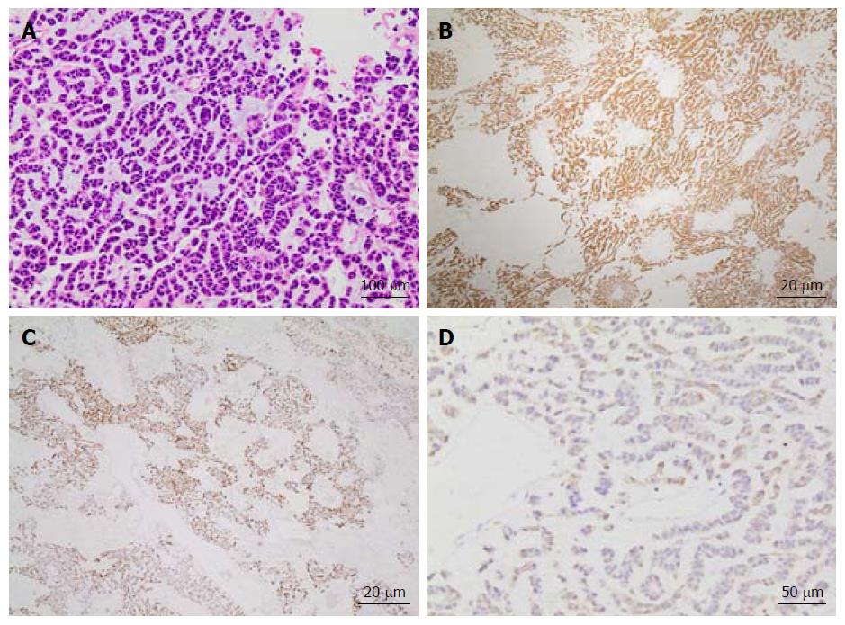
Figure 1 Tissue histopathology of a hepatic lesion confirms metastatic adrenocortical carcinoma.
Hematoxylin and eosin (HE) staining (A) at 200 × magnification of tissue from the hepatic mass shows diffuse infiltrating cords of cells with hyperchromasia, eosinophilic cytoplasm, and a mild degree of nuclear pleomorphism in a background of myxoid stroma. Additional positive immunostaining with inhibin (B) and melan-A (C) at 40 × magnification as well as CKAE1/3 (D) at 100 × magnification confirms the diagnosis as adrenocortical carcinoma.
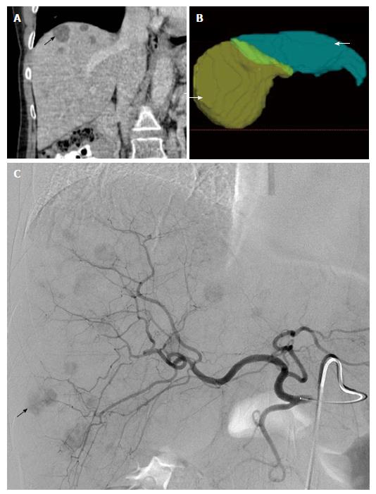
Figure 2 Yttrium-90 planning and radioembolization for treatment of liver metastases from adrenocortical carcinoma.
A: Representative coronal CT abdomen image obtained prior to locoregional therapy shows low density lesions within the liver (arrow) consistent with metastatic disease; B: Three-dimensional reconstructions of the liver used for Yttrium-90 dose calculations revealed a proposed treatment volume of 409 mL for the left hepatic lobe (green colored, arrow) and 1339 mL for the right hepatic lobe (yellow colored, arrowhead) for a total hepatic volume of 1749 mL; C: Digitally subtracted contrast angiography reveals numerous hypervascular metastases within the right and left hepatic lobes (representative right hepatic lobe lesion demarked by arrow).
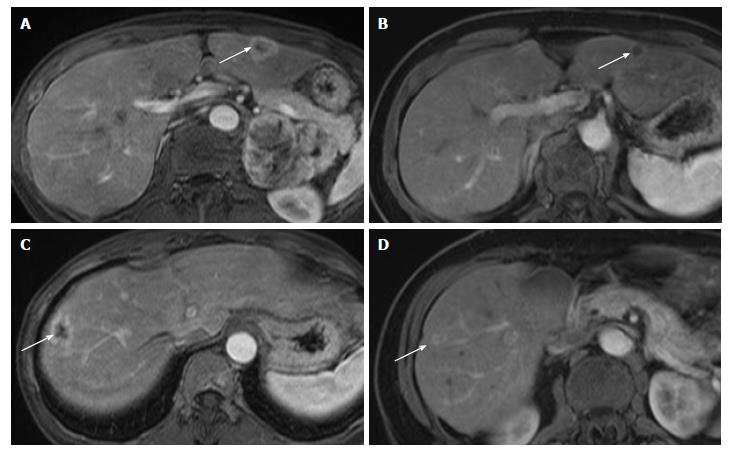
Figure 3 Hepatic metastases from adrenocortical carcinoma respond to Yttrium-90 radioembolization.
Index hepatic lesions within the left hepatic lobe (A) and right hepatic lobe (C) demarked by arrows demonstrate significant reduction in size and perfusion one year following single dose treatment (B and D, respectively, arrows) by catheter-directed Yttrium-90 impregnated microspheres.
- Citation: Makary MS, Krishner LS, Wuthrick EJ, Bloomston MP, Dowell JD. Yttrium-90 microsphere selective internal radiation therapy for liver metastases following systemic chemotherapy and surgical resection for metastatic adrenocortical carcinoma. World J Clin Oncol 2018; 9(1): 20-25
- URL: https://www.wjgnet.com/2218-4333/full/v9/i1/20.htm
- DOI: https://dx.doi.org/10.5306/wjco.v9.i1.20


