Copyright
©The Author(s) 2017.
World J Clin Oncol. Oct 10, 2017; 8(5): 420-424
Published online Oct 10, 2017. doi: 10.5306/wjco.v8.i5.420
Published online Oct 10, 2017. doi: 10.5306/wjco.v8.i5.420
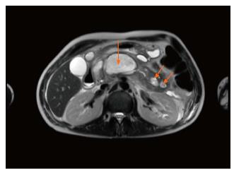
Figure 1 Magnetic resonance imaging of abdomen with and without contrast during July 2016 presentation.
Image displays large walled-off necrosis within the body and tail of the pancreas (arrows).
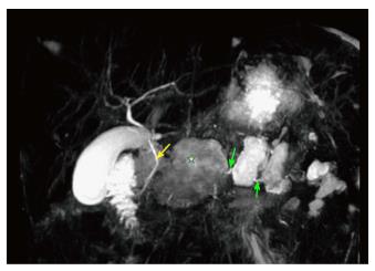
Figure 2 Magnetic resonance cholangiopancreatography performed during July 2016 admission.
Image displays poorly defined main pancreatic duct (green arrows) throughout the pancreas. Common bile duct defined well (yellow arrow) with lack of contrast accentuating the main pancreatic duct. A large walled-off necrosis well imaged again (star).
Figure 3 Endoscopic retrograde cholangiopancreatography performed during July 2016 admission.
Image displays failure of contrast dye to define pancreatic duct and failure of guidewire to cannulate pancreatic duct. Guidewire continues to be diverted to common bile duct which provides evidence of pancreatic duct obstruction.
- Citation: Montminy EM, Landreneau SW, Karlitz JJ. First report of small cell lung cancer with PTHrP-induced hypercalcemic pancreatitis causing disconnected duct syndrome. World J Clin Oncol 2017; 8(5): 420-424
- URL: https://www.wjgnet.com/2218-4333/full/v8/i5/420.htm
- DOI: https://dx.doi.org/10.5306/wjco.v8.i5.420


