Copyright
©The Author(s) 2017.
World J Clin Oncol. Oct 10, 2017; 8(5): 378-388
Published online Oct 10, 2017. doi: 10.5306/wjco.v8.i5.378
Published online Oct 10, 2017. doi: 10.5306/wjco.v8.i5.378
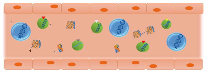
Figure 1 Circulating nucleic acids can be present in different forms.
If actively released, circulating nucleic acids can be found inserted in exosomes (1) and microvesicles (2), or associated with RNA and lipoproteins forming a complex called the virosome (3). Circulating nucleic acids can be also passively released, mainly through apoptosis, in the form of oligo- or mono-nucleosome (4).
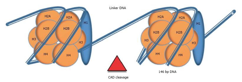
Figure 2 Chromatin cleavage during apoptosis can be a source of circulating DNA.
Circulating DNA can be found in the form of nucleosomes. Each nucleosome is composed of 147 of DNA wrapped around an octamer of histones (H2A, H2B, H3 and H4). An extra histone (H1) stabilizes this complex. During apoptosis, CAD enzyme cleaves in the naked DNA that links each nucleosome (DNA linker), releasing oligo- or mono-nucleosomes. In cancer, where a higher cellular turnover is required, this process can be overloaded and nucleosomes can be secreted into circulation. CAD: Caspase-activated deoxyribonuclease.
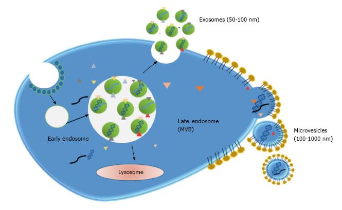
Figure 3 Exosomes and microvesicles can harbour nucleic acids.
Exosomes and microvesicles are generated by different pathways. Exosomes derive from the recycling endosomal pathway, in which the late endosomes (MVB) merge with the plasma membrane instead of with the lysosome, releasing the exosomes. Exosomes encapsulate cytoplasm material such as proteins, RNA or DNA. Microvesicles result from plasma membrane budding, containing cellular components from both cell membrane and cell cytoplasm, including nucleic acids. MVB: Multivesicular bodies.
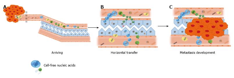
Figure 4 The theory of genometastasis.
The putative mechanism of genometastasis: A: Releasing of nucleic acids to the blood stream; B: Transfection of susceptible cells; C: Malignization of transfected cells.
- Citation: García-Casas A, García-Olmo DC, García-Olmo D. Further the liquid biopsy: Gathering pieces of the puzzle of genometastasis theory. World J Clin Oncol 2017; 8(5): 378-388
- URL: https://www.wjgnet.com/2218-4333/full/v8/i5/378.htm
- DOI: https://dx.doi.org/10.5306/wjco.v8.i5.378


