Copyright
©The Author(s) 2017.
World J Clin Oncol. Feb 10, 2017; 8(1): 91-95
Published online Feb 10, 2017. doi: 10.5306/wjco.v8.i1.91
Published online Feb 10, 2017. doi: 10.5306/wjco.v8.i1.91
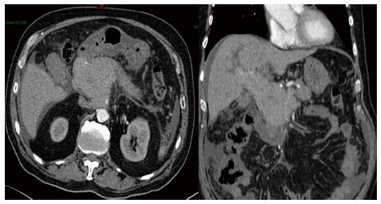
Figure 1 Abdominal computerized tomography scan showing a head pancreas mass extended to the hepatic hilum with mild to moderate dilatation of biliary ducts and a low abundance ascites.
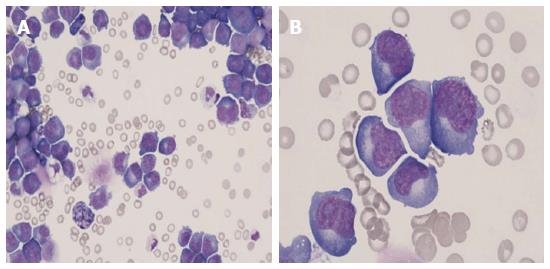
Figure 2 Peritoneal fluid Cytology, May-Grünwald-Giemsa stain.
A: An almost pure population of myeloma cells (× 40); B: Malignant plasma cells exhibiting severe atypia (× 100).
- Citation: Williet N, Kassir R, Cuilleron M, Dumas O, Rinaldi L, Augeul-Meunier K, Cottier M, Roblin X, Phelip JM. Difficult endoscopic diagnosis of a pancreatic plasmacytoma: Case report and review of literature. World J Clin Oncol 2017; 8(1): 91-95
- URL: https://www.wjgnet.com/2218-4333/full/v8/i1/91.htm
- DOI: https://dx.doi.org/10.5306/wjco.v8.i1.91


