Copyright
©The Author(s) 2016.
World J Clin Oncol. Oct 10, 2016; 7(5): 420-424
Published online Oct 10, 2016. doi: 10.5306/wjco.v7.i5.420
Published online Oct 10, 2016. doi: 10.5306/wjco.v7.i5.420
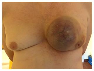
Figure 1 Photodocumentation at time of presentation.
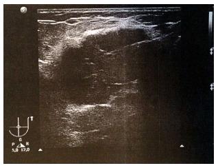
Figure 2 Breast ultrasound shows a large irregular structure of low echogenicity with spiculae measuring > 15 cm × 15 cm (BI-RADS 5).
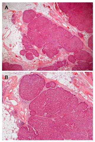
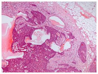
Figure 3 Solid papillary carcinoma (in situ), composed of expansile rounded nodular epithelial masses.
A: Low magnification 12.5 ×; B: Low magnification 25 ×.
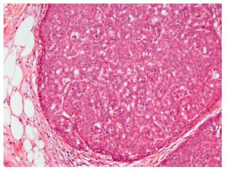
Figure 4 Relatively bland tumor cells with ovoid nuclei and indistinct nucleoli.
Fine fibrovascular septa are seen within the epithelial islands (medium magnification, 100 ×).
Figure 5 Solid papillary carcinoma (invasive) - tumor cell islands with irregular jagged contours within a desmoplastic stroma (medium magnification, 50 ×).
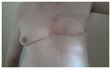
Figure 6 Postoperative clinical presentation.
- Citation: Banys-Paluchowski M, Burandt E, Banys J, Geist S, Sauter G, Krawczyk N, Paluchowski P. Male papillary breast cancer treated by wide resection and latissimus dorsi flap reconstruction: A case report and review of the literature. World J Clin Oncol 2016; 7(5): 420-424
- URL: https://www.wjgnet.com/2218-4333/full/v7/i5/420.htm
- DOI: https://dx.doi.org/10.5306/wjco.v7.i5.420


