Copyright
©The Author(s) 2016.
World J Clin Oncol. Jun 10, 2016; 7(3): 308-320
Published online Jun 10, 2016. doi: 10.5306/wjco.v7.i3.308
Published online Jun 10, 2016. doi: 10.5306/wjco.v7.i3.308
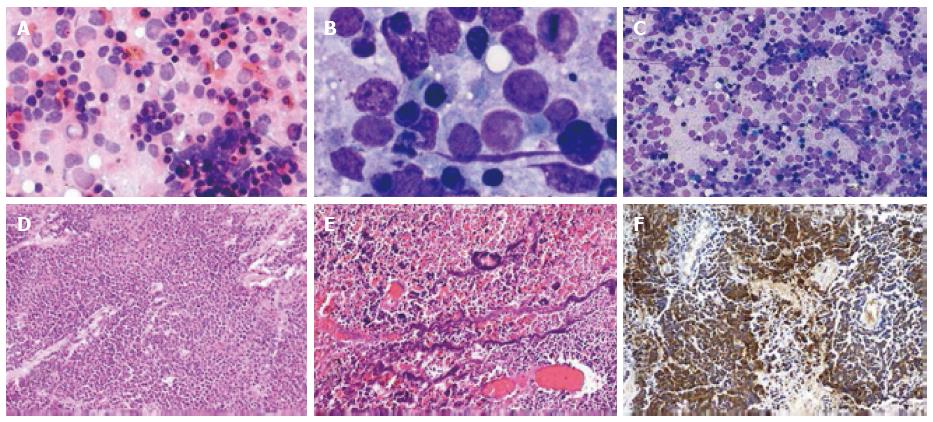
Figure 1 Histopathology and cytology of lymph node samples of illustrative case 1.
A: Microphotograph showing predominantly dispersed population of tumor cells (MGG × 20 ×); B: Microphotograph showing tumor cells with very high nuclear cytoplasmic ratio, scant cytoplasm, round nuclei and fine granular chromatin (MGG × 100 ×); C: Microphotograph showing tumor cells with many apoptotic bodies (HE × 40 ×); D: Photomicrographs showing clusters of tumour cells with small hyperchromatic nuclei, scanty cytoplasm and apoptosis; E: Photomicrographs showing Azzopardi phenomena (basophilic nuclear chromatin spreading to wall of blood vessels); F: Photomicrographs showing synaptophysin immunostain showing intense cytoplasmic positivity.
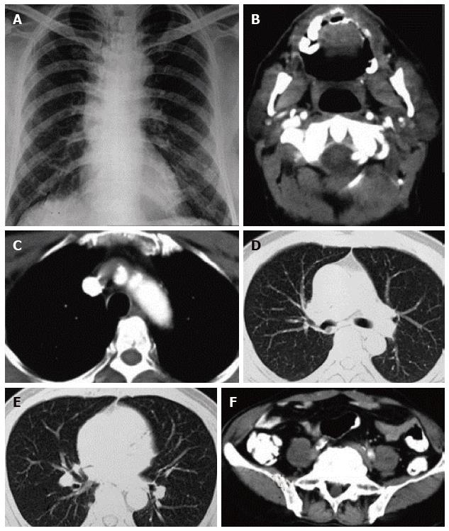
Figure 2 Thoracic imaging at baseline and after treatment of illustrative case 1.
A: Chest radiograph revealing hyperinflated lung fields with no evidence of any parenchymal abnormality; B: Contrast enhanced computed tomography (CECT) of the neck revealing enlarged right sided cervical group of lymph nodes; C: Mediastinal window of CECT of the thorax with no evidence of mediastinal lymph node enlargement; D and E: Lung window of CECT thorax with no evidence of primary in the lung; F: CECT of the abdomen with no evidence of any abnormality in the abdomen.
Figure 3 Histopathology and cytology of lymph node samples of illustrative case 2.
A: Microphotograph showing dispersed population of tumor cells along with few loose clusters. The tumor cells with high nuclear cytoplasmic ratio and showing nuclear moulding (MGG × 20 ×); B: Microphotograph showing small tumor cells with high N:C ratio, salt and pepper type chromatin and inconspicuous nucleoli (HE × 40 ×); C: Photomicrographs showing tumour with extensive crushing artefact; D: Tumour cells having hyperchromatic nuclei, scanty cytoplasm and apoptosis; E: Synaptophysin immunostain showing cytoplasmic positivity.
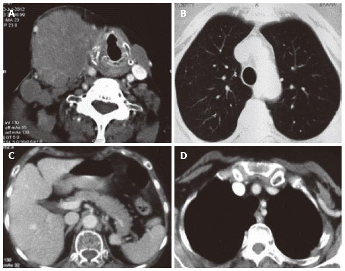
Figure 4 Thoracic imaging at baseline and after treatment of case 2.
A: Contrast enhanced computed tomography of the neck revealing a conglomerate lymph node mass of size 7 cm × 4 cm involving the right submandibular region. The mass is pushing the larynx towards the left side; B: Contrast enhanced computed tomography of the thorax (lung window) with no evidence of primary in the lung; C: Contrast enhanced computed tomography of the abdomen with normal abdominal organs and no evidence of any primary in the abdomen; D: Mediastinal window of CECT thorax revealing no enlarged lymph node stations in the mediastinum. CECT: Contrast enhanced computed tomography.
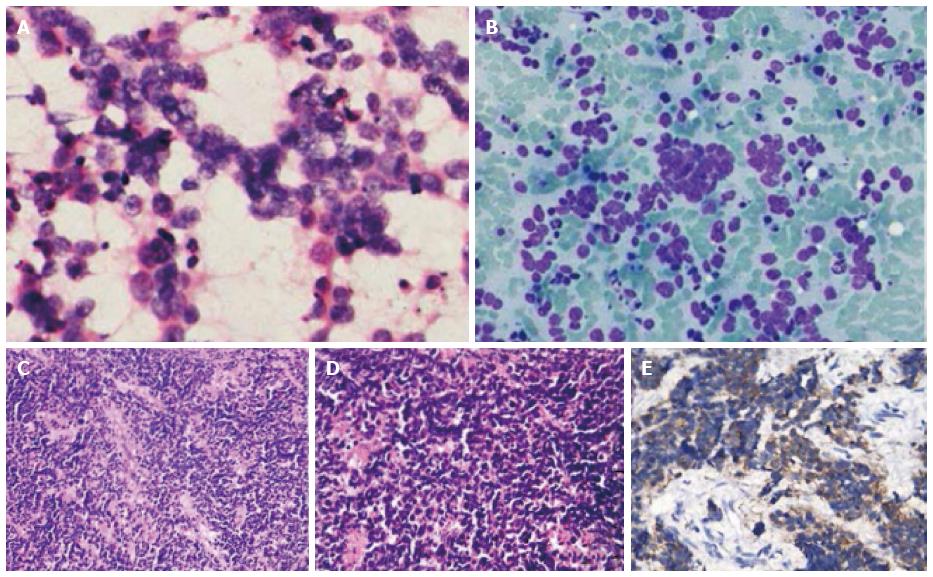
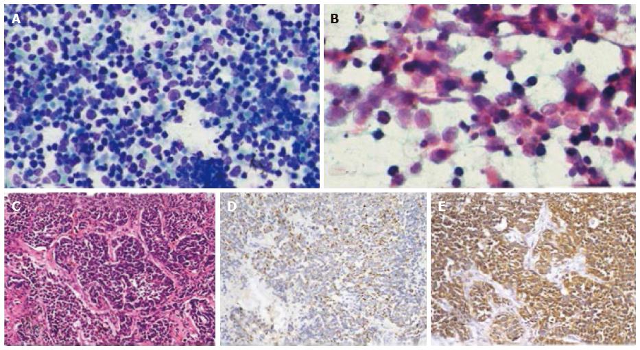
Figure 5 Histopathology and cytology of lymph node samples of case 3.
A: Microphotograph showing dispersed population of small sized tumor cells with high nuclear cytoplasmic ratio along with many degenerated cells (MGG × 20 ×); B: Microphotograph showing nuclear threading and crushing along with tumor cells with round nuclei, high N:C ratio, salt and pepper type chromatin and inconspicuous nucleoli (Pap × 40 ×); C: Photomicrographs showing clusters of tumour cells with hyperchromatic nuclei, nuclear molding and scanty cytoplasm; D: Pancytokeratin staining showing patchy dot like positivity; E: Synaptophysin immunostain showing intense cytoplasmic positivity.
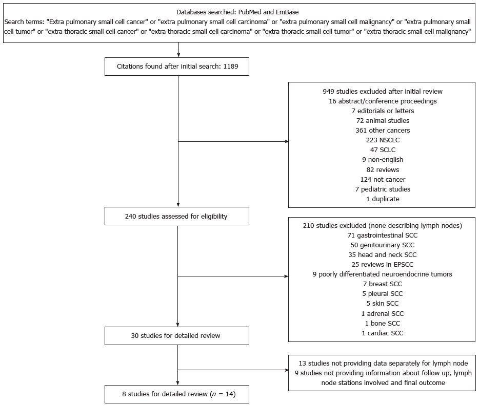
Figure 6 Study selection process for systematic review.
NSCLC: Non small cell lung cancer; SCLC: Small cell lung cancer; SCC: Small cell cancer; EPSCC: Extrapulmonary small cell cancer.
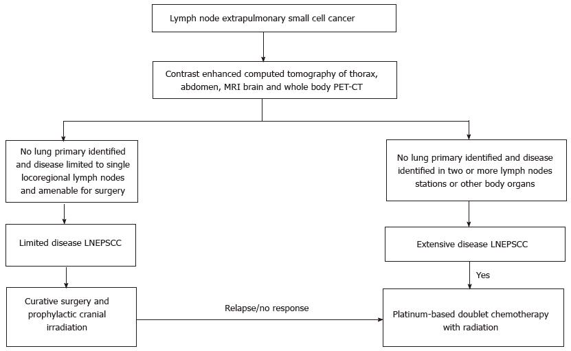
Figure 7 Algorithm for the diagnostic work-up and treatment of lymph node extrapulmonary small cell carcinoma.
LNEPSCC: Lymph nodes extrapulmonary small cell carcinoma; PET: Positron emission tomography; CT: Computed tomography; MRI: Magnetic resonance imaging.
- Citation: Sehgal IS, Kaur H, Dhooria S, Bal A, Gupta N, Behera D, Singh N. Extrapulmonary small cell carcinoma of lymph node: Pooled analysis of all reported cases. World J Clin Oncol 2016; 7(3): 308-320
- URL: https://www.wjgnet.com/2218-4333/full/v7/i3/308.htm
- DOI: https://dx.doi.org/10.5306/wjco.v7.i3.308


