Copyright
©The Author(s) 2016.
World J Clin Oncol. Apr 10, 2016; 7(2): 270-274
Published online Apr 10, 2016. doi: 10.5306/wjco.v7.i2.270
Published online Apr 10, 2016. doi: 10.5306/wjco.v7.i2.270
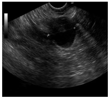
Figure 1 Ultrasound of the right ovary showing a smooth cyst with intracystic papillary structures.
There was no ascites.
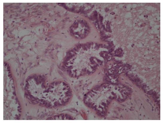
Figure 2 Borderline tumor of the right ovary.
Hyperchromatic nuclei with large nucleoli; tumorous structures with hypervascularized papillary stroma without invasion into the center of the cyst.
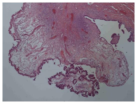
Figure 3 Epifascial non-invasive implant of a papillary borderline tumor.
- Citation: Banys-Paluchowski M, Yeganeh B, Luettges J, Maibach A, Langenberg R, Krawczyk N, Paluchowski P, Maul H, Gebauer G. Isolated subcutaneous implantation of a borderline ovarian tumor: A case report and review of the literature. World J Clin Oncol 2016; 7(2): 270-274
- URL: https://www.wjgnet.com/2218-4333/full/v7/i2/270.htm
- DOI: https://dx.doi.org/10.5306/wjco.v7.i2.270


