Copyright
©The Author(s) 2015.
World J Clin Oncol. Dec 10, 2015; 6(6): 225-236
Published online Dec 10, 2015. doi: 10.5306/wjco.v6.i6.225
Published online Dec 10, 2015. doi: 10.5306/wjco.v6.i6.225
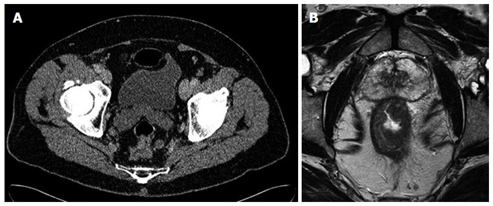
Figure 1 Computed tomography (A) and magnetic resonance imaging (B) of the T3 rectal cancer.
Note poor quality of circumferential margin on the computed tomography scan compared to the magnetic resonance imaging.
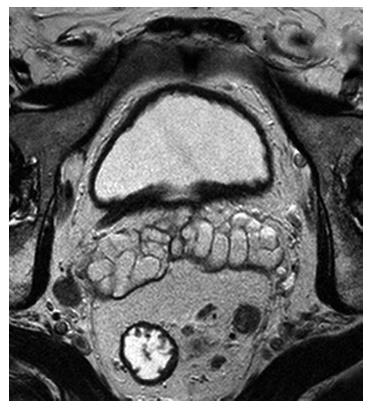
Figure 2 Magnetic resonance imaging of the T3 rectal cancer showing lymph node involvement.
- Citation: Milinis K, Thornton M, Montazeri A, Rooney PS. Adjuvant chemotherapy for rectal cancer: Is it needed? World J Clin Oncol 2015; 6(6): 225-236
- URL: https://www.wjgnet.com/2218-4333/full/v6/i6/225.htm
- DOI: https://dx.doi.org/10.5306/wjco.v6.i6.225


