Copyright
©The Author(s) 2015.
World J Clin Oncol. Aug 10, 2015; 6(4): 57-63
Published online Aug 10, 2015. doi: 10.5306/wjco.v6.i4.57
Published online Aug 10, 2015. doi: 10.5306/wjco.v6.i4.57
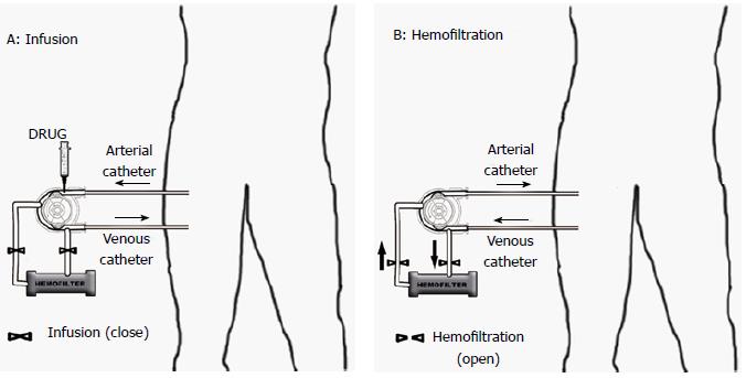
Figure 1 Limb infusion/hemofiltration scheme.
A: Infusion: the blood is drawn from the venous line and, once enriched with the drug, is reintroduced in the arterial line. The hemofilter is excluded from the circuit. The occlusion of the blood inflow and outflow was achieved by catheters with terminal ballon inflated; B: Hemofiltration: lines are reversed: the blood is drawn from the arterial side and reintroduces into the venous line. The hemofilter is connected to the circuit.
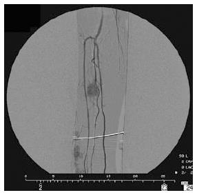
Figure 2 Angiogram of melanoma.
In a patient with extremity melanoma AJCC stage IIIC, angiogram shows in the middle third of the leg rounded nodule with a homogeneous, hypervascular stain, nourished by muscular branches of the peroneal artery. Additional deep-seated lesions, which were not palpable clinically, were detected by angiography distally in the leg, nourished by branches of the anterior tibial artery.
- Citation: Cecchini S, Sarti D, Ricci S, Vergini LD, Sallei M, Serresi S, Ricotti G, Mulazzani L, Lattanzio F, Fiorentini G. Isolated limb infusion chemotherapy with or without hemofiltration for recurrent limb melanoma. World J Clin Oncol 2015; 6(4): 57-63
- URL: https://www.wjgnet.com/2218-4333/full/v6/i4/57.htm
- DOI: https://dx.doi.org/10.5306/wjco.v6.i4.57


