Copyright
©2014 Baishideng Publishing Group Inc.
World J Clin Oncol. Dec 10, 2014; 5(5): 982-989
Published online Dec 10, 2014. doi: 10.5306/wjco.v5.i5.982
Published online Dec 10, 2014. doi: 10.5306/wjco.v5.i5.982
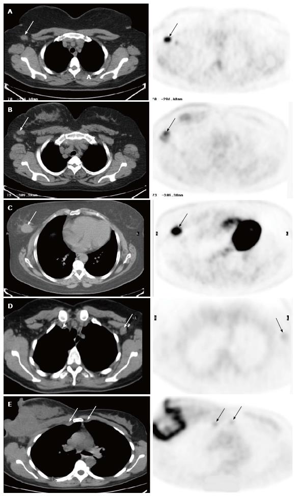
Figure 1 Fluorodeoxyglucose positron emission tomography/computed tomography.
A: In a 61-year-old woman with newly diagnosed breast cancer. There was a single palpable axillary lymph node on physical examination. The images demonstrated a 1.5 cm axillary node with intense uptake (SUV 10, arrows), consistent with metastasis. There were also additional smaller axillary nodes with abnormal uptake. The patient underwent axillary lymph node dissection (ALND), and surgical pathology revealed that two axillary nodes were positive; B: In a 51-year-old woman with newly diagnosed right breast cancer. There were palpable right axillary lymph nodes on physical examination. The images demonstrated a few enlarged right axillary nodes with intense uptake (SUV 6.5, arrows). The patient had ALND, which showed three metastatic lymph nodes; C: In a 54-year-old woman with newly diagnosed right breast cancer. The images showed a 3.0 cm fluorodeoxyglucose (FDG) avid tumor in the right breast (arrows), but were negative in the right axilla. The patient had lymphscintigraphy for sentinel lymph node biopsy, which was negative for axillary metastasis; D: In a 40-year-old woman with history of left breast cancer and post lumpectomy. On physical examination, there was a palpable lymph node in the left axilla. Positron emission tomography/computed tomography (PET/CT) showed a 1.5 cm left axillary lymph node with very mild uptake (SUV 1.4, arrows), consistent with a benign etiology. Subsequent SLNB confirmed lymphadenitis; E: FDG PET/CT in a 41-year-old woman with newly diagnosed inflammatory carcinoma of the right breast. In addition to large right breast necrotic mass and axillary nodal lesions, there are a 1.2 cm right internal mammary node (IMN) and a 1.0 cm left IMN, and both are mildly FDG avid (SUV 3.2) and suspicious for IMN metastases (arrows). The patient was excluded as a surgical candidate based on FDG PET/CT findings. She received chemotherapy and radiation. SUV: Standardized uptake value.
- Citation: Liu Y. Role of FDG PET-CT in evaluation of locoregional nodal disease for initial staging of breast cancer. World J Clin Oncol 2014; 5(5): 982-989
- URL: https://www.wjgnet.com/2218-4333/full/v5/i5/982.htm
- DOI: https://dx.doi.org/10.5306/wjco.v5.i5.982


