Copyright
©2014 Baishideng Publishing Group Inc.
World J Clin Oncol. Dec 10, 2014; 5(5): 921-930
Published online Dec 10, 2014. doi: 10.5306/wjco.v5.i5.921
Published online Dec 10, 2014. doi: 10.5306/wjco.v5.i5.921
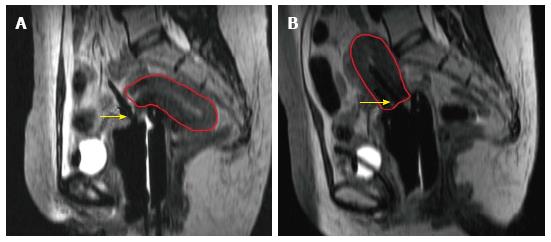
Figure 1 Uterine perforation despite perfect geometric applicator placement relative to bone anatomy.
A: Shows the uterine perforation with retroverted uterus (red) diagnosed on magnetic resonance imaging (MRI) imaging for brachytherapy planning; B: Shows appropriate tandem placement (yellow arrow) ameliorated by placing the applicator under ultra-sound guidance and confirmed by MRI image-based planning.
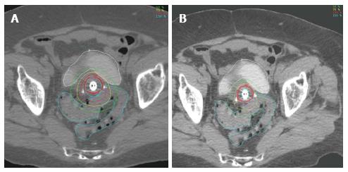
Figure 2 Conventional 2D point dose brachytherapy planning over doses critical organs for small cervix.
Magnetic resonance imaging planning images showing over-dosage of critical organs with point-A image based prescription (A) for patient with small cervix which is improved with image-based brachytherapy (B).
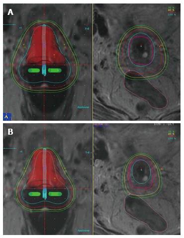
Figure 3 Sigmoid colon sparing with image-based planning.
Magnetic resonance imaging planning images showing point A optimized plan with high doses to adjacent sigmoid colon (A), which is reduced with image-based 3D optimization (B).
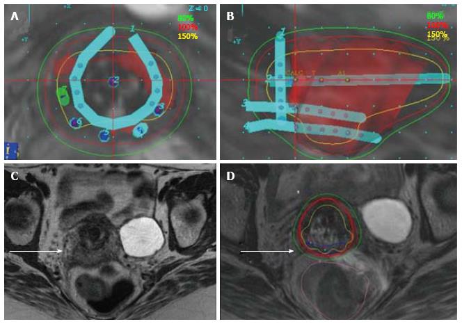
Figure 4 Use of the vienna applicator to improve posterior disease coverage for locally-advanced cervical cancer.
A and B: 3D reconstruction of Vienna applicator showing posterior needles; C and D axial magnetic resonance imaging (MRI) showing advanced disease with posterior extension, and improved coverage with combined interstitial and intracavitary MRI image-based planning.
- Citation: Vargo JA, Beriwal S. Image-based brachytherapy for cervical cancer. World J Clin Oncol 2014; 5(5): 921-930
- URL: https://www.wjgnet.com/2218-4333/full/v5/i5/921.htm
- DOI: https://dx.doi.org/10.5306/wjco.v5.i5.921


