Copyright
©2014 Baishideng Publishing Group Inc.
World J Clin Oncol. Dec 10, 2014; 5(5): 1113-1116
Published online Dec 10, 2014. doi: 10.5306/wjco.v5.i5.1113
Published online Dec 10, 2014. doi: 10.5306/wjco.v5.i5.1113
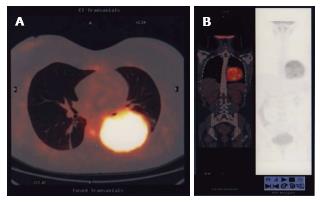
Figure 1 Computed tomography/positron emission tomography scan.
A: CT-PET scan showing the large round mass in the left basal hemithorax; B: CT-PET scan showing a frontal vision of the mass inside the chest. CT-PET: Computed tomography/positron emission tomography.
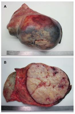
Figure 2 A left posterolateral thoracotomy with lower lobectomy.
A: Macroscopic vision of the large tumor infiltrating the left lower lobe; B: The cutting surface of the large tumor inside the left lower lobe.
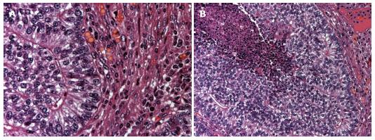
Figure 3 Monophasic pulmonary blastoma.
A: Microscopic appearance of cytology in monophasic pulmonary blastoma; B: Monophasic pulmonary blastoma: Prevalence of epithelial on mesenchymal elements.
- Citation: Magistrelli P, D’Ambra L, Berti S, Bonfante P, Francone E, Vigani A, Falco E. Adult pulmonary blastoma: Report of an unusual malignant lung tumor. World J Clin Oncol 2014; 5(5): 1113-1116
- URL: https://www.wjgnet.com/2218-4333/full/v5/i5/1113.htm
- DOI: https://dx.doi.org/10.5306/wjco.v5.i5.1113


