Copyright
©2014 Baishideng Publishing Group Inc.
World J Clin Oncol. Dec 10, 2014; 5(5): 1107-1112
Published online Dec 10, 2014. doi: 10.5306/wjco.v5.i5.1107
Published online Dec 10, 2014. doi: 10.5306/wjco.v5.i5.1107
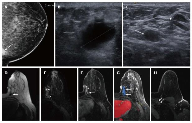
Figure 1 Imaging findings.
A: Medially exaggerated cranio-caudal mammographic view of the left breast demonstrates the anterior aspect of the palpable mass that is a partially circumscribed and partially indistinct mass at 9 o’clock, posterior left breast (arrow); B: Ultrasound of the palpable lump demonstrates a 2.1 cm indistinct irregular hypoechoic solid mass with no posterior shadowing (caliper); C: Ultrasound of the left axilla demonstrates multiple oval and rounded lymph nodes measuring up to 1.4 cm. One oval 1.1 cm lymph node is diffusely hypoechoic (arrow) and is targeted for ultrasound guided Fine Needle Aspiration. The majority of the remaining lymph nodes demonstrate a thickened cortex (arrow head); D-H: Breast MRI: axial images of the left breast shown in multiple series as below; D: On pre-contrast T1W non-fat suppressed imaging the palpable mass is a 2 cm irregular mass with a central artifact from clip (arrow); E: On pre-contrast T2W fat suppressed imaging the mass is isointense to slightly hyperintense relative to the breast parenchyma (arrow). Similar signal is noted anterior to the mass with a higher intensity area more anteriorly (arrowhead); F: On first post-contrast fat suppressed T1W imaging the 2 cm irregular mass with a clip (arrow) is enhancing. Additional non-mass enhancement is noted anterior to the mass, measuring approximately 3 cm in the anterior-posterior plane (open arrow); G: Computerized color mapping demonstrates mostly persistent kinetic pattern of the mass and non-mass enhancement (area in blue, arrows); H: First post-contrast fat suppressed T1W imaging confirms the presence of multiple abnormal level 1 (arrow) and level 2 (not shown) axillary lymph nodes and abnormal left internal mammary chain adenopathy (double arrows).
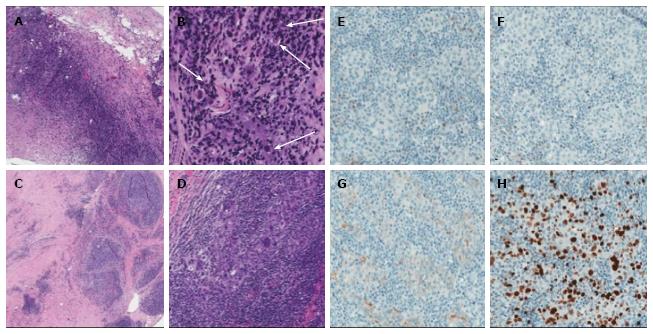
Figure 2 Pathology findings.
A, B: Breast, core needle biopsy: large area of necrosis with chronic inflammation and fibrosis at periphery of lesion, 4X magnification (A); 16X magnification of A showing rare, atypical cells (arrows, B); C, D: Breast, surgical excision: poorly circumscribed lesion adjacent to previous biopsy site, 10X magnification (C); high grade carcinoma with marked pleomorphism and syncytical growth somewhat obscured by marked intra- and peri-turmoral inflammatory infiltrate (D) characteristic of LELC, 10X magnification; E-H: Immunophenotype: ER negative (E), PR negative (F), Her-2/neu 1+ (G), and Ki-67 27% (H). 10X magnification. ER: Estrogen-receptor; PR: Progesterone-receptor.
- Citation: Suzuki I, Chakkabat P, Goicochea L, Campassi C, Chumsri S. Lymphoepithelioma-like carcinoma of the breast presenting as breast abscess. World J Clin Oncol 2014; 5(5): 1107-1112
- URL: https://www.wjgnet.com/2218-4333/full/v5/i5/1107.htm
- DOI: https://dx.doi.org/10.5306/wjco.v5.i5.1107


