Copyright
©2014 Baishideng Publishing Group Inc.
World J Clin Oncol. Aug 10, 2014; 5(3): 224-240
Published online Aug 10, 2014. doi: 10.5306/wjco.v5.i3.224
Published online Aug 10, 2014. doi: 10.5306/wjco.v5.i3.224
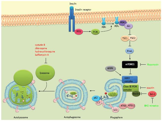
Figure 1 The autophagic process and some of its major regulators.
The phosphatidylinositol 3-kinase (PI3K) pathway is triggered by the binding of insulin or growth factors to the insulin receptor, activating PI3K. Activated PI3K converts PIP2 to PIP3 which then recruits PDK1 and Akt to the plasma membrane. Activated Akt then phosphorylates and inactivates TSC 1/2, leading to the activation of Rheb and mammalian target of rapamycin complex 1 (mTORC1). Under nutrient-rich conditions, mTORC1 suppresses autophagy by phosphorylating and inhibiting the ULK1 complex. Under starvation conditions or rapamycin treatment, mTOR dissociates from the ULK1 complex and autophagy gets activated. ULK1 is also directly phosphorylated and activated by the AMP-activated protein kinase (AMPK) in response to energy restriction. The autophagic process involves the degradation of cytosolic proteins and organelles in the lysosomes via autophagosomal delivery. Two complexes regulate the formation of the phagophore, the ULK1 complex and the beclin 1-VPS34 (class III PI3K) complex. The elongation of the autophagosomal membrane is mediated by two ubiquitin-like protein conjugation systems: ATG12-ATG5 and ATG8/LC3. LC3 can additionally recruit adaptor proteins such as p62 to autophagosomes mediating selective autophagy of cellular structures, protein aggregates and microorganisms. LC3II (LC3 bound to phosphatidylethanolamine) is recruited to both the inner and outer autophagosomal membrane. Autophagosomes fuse with lysosomes to form autolysosomes where they are degraded together with their luminal content. Pharmacological modulators of autophagy are labeled according to their effect on autophagy. Red: Inhibits autophagy; Green: Induces autophagy.
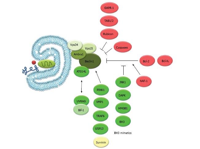
Figure 2 Beclin 1, its protein interactions and its role in autophagy.
Beclin 1 has been found to form part of different class III phosphatidylinositol 3-kinase (PI3K) complexes. Each complex consists of beclin 1, Vps34, Vps15 and Ambra1. ATG14L activates the complex and induces the formation of autophagosomes. UVRAG and ATG14L are present in mutually exclusive class III PI3K complexes and UVRAG and Bif-1 have been shown to activate the complex. UVRAG has also been shown to function in autophagosome maturation and endocytic trafficking possibly independent from its interaction with beclin 1. Rubicon has also been shown to bind beclin 1 and can inhibit autophagosome formation and maturation. TAB1/2, two upstream activators of the TAK1-IKK signaling pathway interact with beclin 1 and their dissociation seems to be necessary for autophagy induction. GAPR-1 can also bind beclin 1 and inhibit autophagy probably through beclin 1 tethering in the Golgi apparatus. Bcl-2/Bcl-XL can bind beclin 1 and inhibit autophagy. JNK1 phosphorylates Bcl-2 while DAPK phosphorylates beclin 1 and disrupt their interaction. Additionally, BH3-only proteins (tBid, Bad, BNIP3) or BH3 mimetics (ABT737) bind Bcl-2 and release beclin 1. Beclin 1 has also been found to be a substrate of caspases-3, 7 and 8 during apoptosis, in which cleavage of beclin 1 suppresses autophagy. Other beclin 1 interacting proteins that induce autophagy are PINK1 and VMP1. TRAF6 and USP13 have been shown to regulate beclin 1 ubiquitination. Survivin, an anti-apoptotic protein can also bind beclin 1 and regulate TRAIL induced apoptosis. NAF-1, a component of the IP3 receptor complex contributes to the interaction of Bcl-2 with beclin 1 at the ER[32,99-104]. Proteins and drugs shown are color-coded according to their effect on autophagy. Red: Inhibits autophagy; Green: Induces autophagy; Yellow: Unknown.
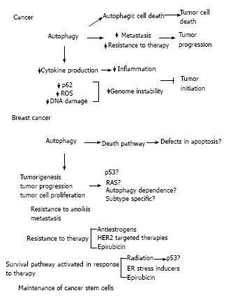
Figure 3 Autophagy in cancer and breast cancer.
Different roles have been described for autophagy in cancer. Autophagy can limit tumor initiation by decreasing inflammation and genome instability. Once a tumor is established, autophagy can induce tumor progression by facilitating metastasis and resistance to therapy. On the other hand, it has also been proposed that autophagy could function as a cell death process that could be induced during therapy. In breast cancer, although some evidence suggests that autophagic cell death occurs in apoptosis defective cells, most of the evidence in the literature suggests a tumor promoting role for autophagy. Autophagy has been shown to promote tumorigenesis, tumor proliferation and progression. This could depend on the p53 or RAS mutation status of the cells. Also, if breast cancer is an autophagy-dependent disease remains to be determined as well as if this dependence is subtype specific. Autophagy has also been involved in resistance to anoikis, facilitation of metastasis, resistance to therapy and maintenance of breast cancer stem cells. Moreover, when cancer cells are made resistant to the therapies shown in the figure, they show increased autophagy and inhibition of autophagy reverts this resistance. Therapies that are known to induce autophagy in breast cancer are also shown. With these treatments, autophagy inhibition has been shown to increase cell death. Regarding radiation, results are controversial and the role of autophagy could depend on the p53 status of the cell.
- Citation: Maycotte P, Thorburn A. Targeting autophagy in breast cancer. World J Clin Oncol 2014; 5(3): 224-240
- URL: https://www.wjgnet.com/2218-4333/full/v5/i3/224.htm
- DOI: https://dx.doi.org/10.5306/wjco.v5.i3.224


