Copyright
©2012 Baishideng Publishing Group Co.
World J Clin Oncol. Dec 10, 2012; 3(12): 155-158
Published online Dec 10, 2012. doi: 10.5306/wjco.v3.i12.155
Published online Dec 10, 2012. doi: 10.5306/wjco.v3.i12.155
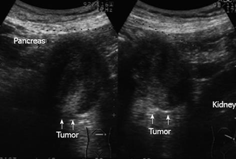
Figure 1 Abdominal ultrasonography showed a low-echoic mass sandwiched between the pancreas and left kidney, measuring 55 mm in its greatest diameter (arrows).
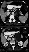
Figure 2 Abdominal computed tomography demonstrated a unilocular cystic tumor that appeared to be situated in the pancreas and contact with the left adrenal gland (arrowhead).
The tumor had a thin wall with calcification, and a solid component was apparent (arrow).
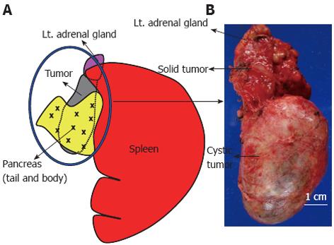
Figure 3 The schema shows the macroscopic appearance of the resected specimen.
A: The cystic tumor (circled in blue) was separated from the pancreas by a fibrous tumor membrane; B: The tumor measured 10 cm × 4 cm in diameter and was filled with homogenous, grayish, creamy fluid. This tumor also had a solid component measuring 3 cm × 2.5 cm in diameter adjacent to the left adrenal gland.
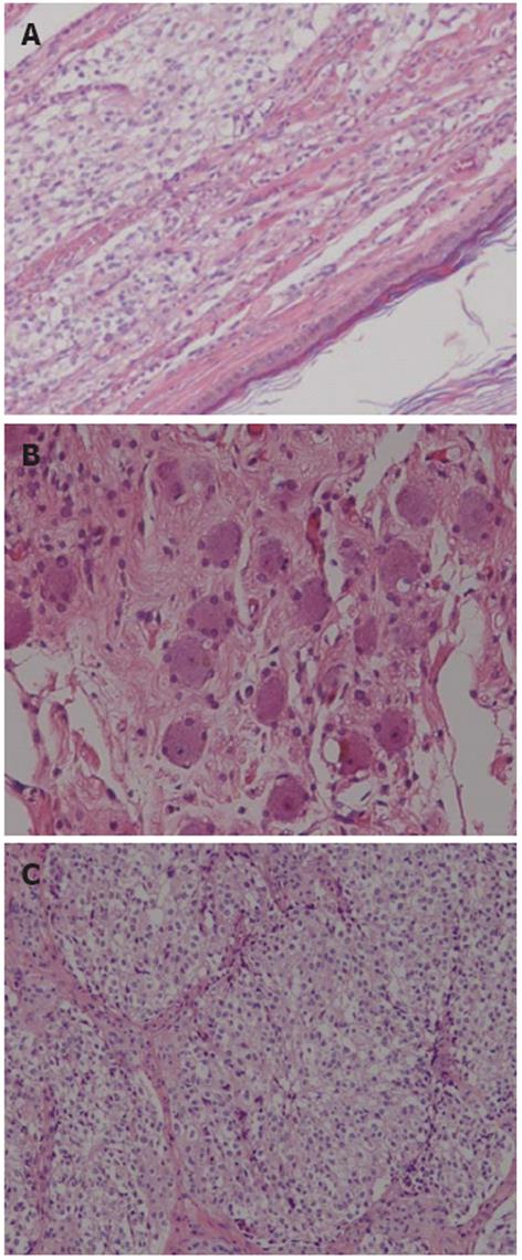
Figure 4 Histopathologic findings of the tumor.
A: Benign squamous epithelium surrounding the cystic area (right lower part of figure) and ganglioneuroblastoma components (left upper part of figure) were adjacent and closely apposed to each other; B: Microscopic view of the mature, benign part of the ganglioneuroblastoma, showing ganglion cells and nerve fibers (hematoxylin and eosin, × 200); C: the malignant part of the ganglioneuroblastoma composed of neuroblasts (hematoxylin and eosin, × 200).
- Citation: Hayama S, Ohmi M, Yonemori A, Yamabuki T, Inomata H, Nihei K, Hirano S. Ganglioneuroblastoma arising within a retroperitoneal mature cystic teratoma. World J Clin Oncol 2012; 3(12): 155-158
- URL: https://www.wjgnet.com/2218-4333/full/v3/i12/155.htm
- DOI: https://dx.doi.org/10.5306/wjco.v3.i12.155


