Copyright
©2012 Baishideng Publishing Group Co.
World J Clin Oncol. Jan 10, 2012; 3(1): 12-14
Published online Jan 10, 2012. doi: 10.5306/wjco.v3.i1.12
Published online Jan 10, 2012. doi: 10.5306/wjco.v3.i1.12
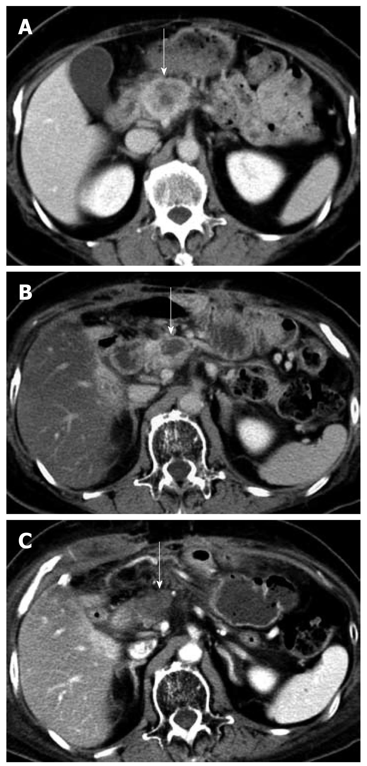
Figure 1 Abdominal contrast-enhanced computed tomography scan.
A: Contrast-enhanced computed tomography (CE-CT) scan showing a tumor in the pancreatic head before the first laparotomy (arrow); B: CE-CT scan taken after the chemoradiotherapy showing a reduction in the size of the tumor (arrow); C: CE-CT scan taken after the radiofrequency ablation showing a necrotic area that is suggestive of an ablated tumor (arrow).
- Citation: Ikuta S, Kurimoto A, Iida H, Aihara T, Takechi M, Kamikonya N, Yamanaka N. Optimal combination of radiofrequency ablation with chemoradiotherapy for locally advanced pancreatic cancer. World J Clin Oncol 2012; 3(1): 12-14
- URL: https://www.wjgnet.com/2218-4333/full/v3/i1/12.htm
- DOI: https://dx.doi.org/10.5306/wjco.v3.i1.12


