Copyright
©2010 Baishideng Publishing Group Co.
World J Clin Oncol. Feb 10, 2011; 2(2): 94-107
Published online Feb 10, 2011. doi: 10.5306/wjco.v2.i2.94
Published online Feb 10, 2011. doi: 10.5306/wjco.v2.i2.94
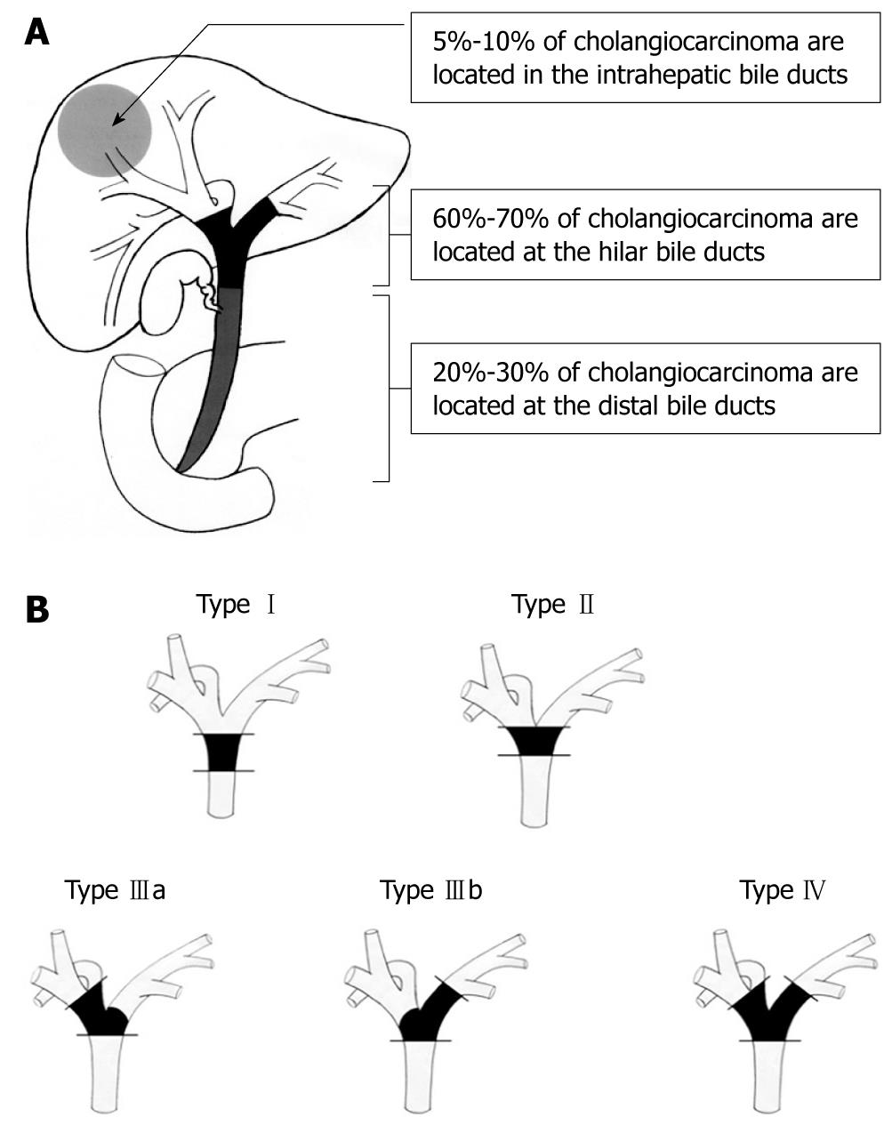
Figure 1 Anatomic classification of cholangiocarcinoma.
A: The majority of cholangiocarcinoma (60%-70%) develop in the hilar bile duct and are called Klatskin tumors. The distal bile duct is involved in 20% to 30% of cases, while intrahepatic cholangiocarcinomas represent 5% to 10% of the tumors originating from the biliary tract; B: Bismuth-Corlette classification of hilar bile duct cancer. Type I, cholangiocarcinoma confined to the common bile duct; Type II, cholangiocarcinoma involves the bifurcation of the common bile duct; Type IIIa, cholangiocarcinoma involves the bifurcation and the right hepatic duct; Type IIIb, cholangiocarcinoma involves the bifurcation and the left hepatic duct; Type IV, cholangiocarcinoma involves the bifurcation and extends to both the right and left hepatic ducts.
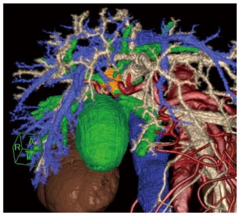
Figure 2 Hilar cholangiocarcinoma (Bismuth-Corlette type IIIa).
Comprehensive multiphase fusion images of the tumor (orange), bile duct (green), and surrounding vessels including hepatic artery (red), portal vein (light yellow), and hepatic vein (blue).
- Citation: Akamatsu N, Sugawara Y, Hashimoto D. Surgical strategy for bile duct cancer: Advances and current limitations. World J Clin Oncol 2011; 2(2): 94-107
- URL: https://www.wjgnet.com/2218-4333/full/v2/i2/94.htm
- DOI: https://dx.doi.org/10.5306/wjco.v2.i2.94


