Copyright
©2010 Baishideng Publishing Group Co.
World J Clin Oncol. Feb 10, 2011; 2(2): 108-114
Published online Feb 10, 2011. doi: 10.5306/wjco.v2.i2.108
Published online Feb 10, 2011. doi: 10.5306/wjco.v2.i2.108
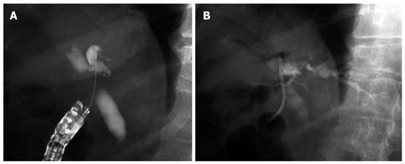
Figure 1 Endoscopic ultrasonography biliary drainage using the rendezvous technique.
A: Intrahepatic approach: fluoroscopic image of the wire crossing the hilar stricture and advancing into the duodenum; B, C: Subsequent endoscopic retrograde cholangiopancreatography with bile duct access and plastic stent placement.
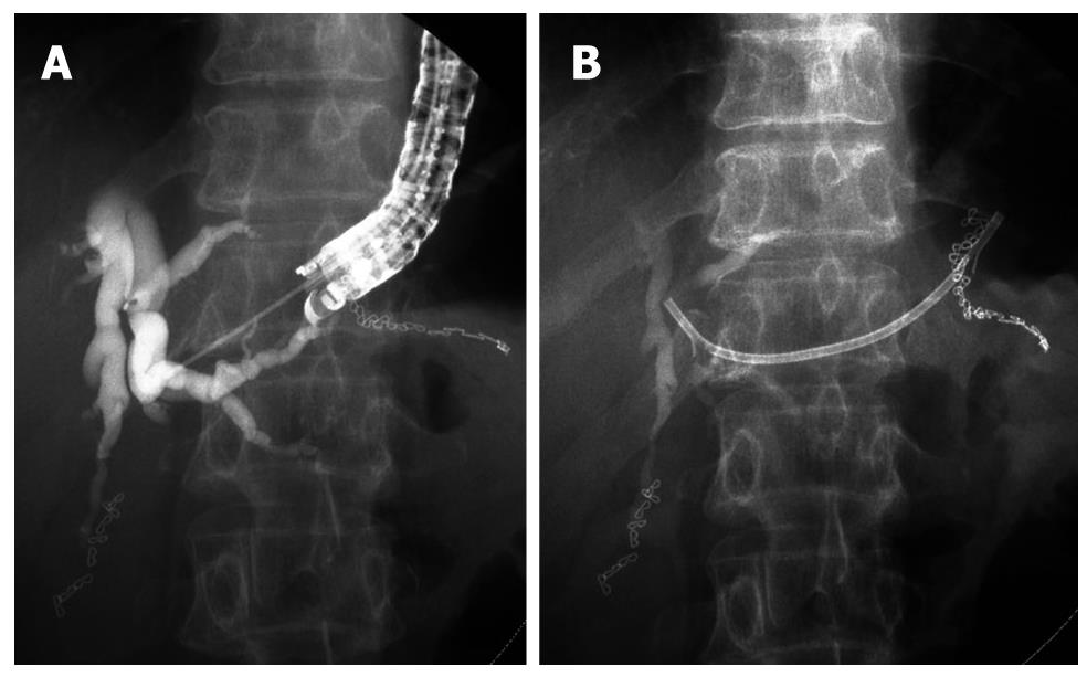
Figure 2 Endoscopic ultrasonography-guided choledochoduodenostomy.
A: Cholangiography after puncture of extrahepatic bile duct by needle knife; B: Plastic stent was inserted from duodenum into extrahepatic bile duct.
Figure 3 Endoscopic ultrasonography-guided hepaticogastrostomy.
A: Cholangiography after puncture of intrahepatic bile duct by 19G fine needle. B: Plastic stent was inserted from stomach into intrahepatic bile duct.
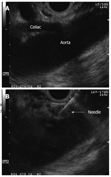
Figure 4 Endoscopic ultrasonography-guided celiac plexus neurolysis.
A: At first, we visualize the celiac trunk by linear array echoendoscope; B: Endoscopic ultrasonography image during ethanol injection.
- Citation: Hara K, Yamao K, Mizuno N, Hijioka S, Sawaki A, Tajika M, Kawai H, Kondo S, Shimizu Y, Niwa Y. Interventional endoscopic ultrasonography for pancreatic cancer. World J Clin Oncol 2011; 2(2): 108-114
- URL: https://www.wjgnet.com/2218-4333/full/v2/i2/108.htm
- DOI: https://dx.doi.org/10.5306/wjco.v2.i2.108


