Copyright
©2011 Baishideng Publishing Group Co.
World J Clin Oncol. Oct 10, 2011; 2(10): 348-354
Published online Oct 10, 2011. doi: 10.5306/wjco.v2.i10.348
Published online Oct 10, 2011. doi: 10.5306/wjco.v2.i10.348
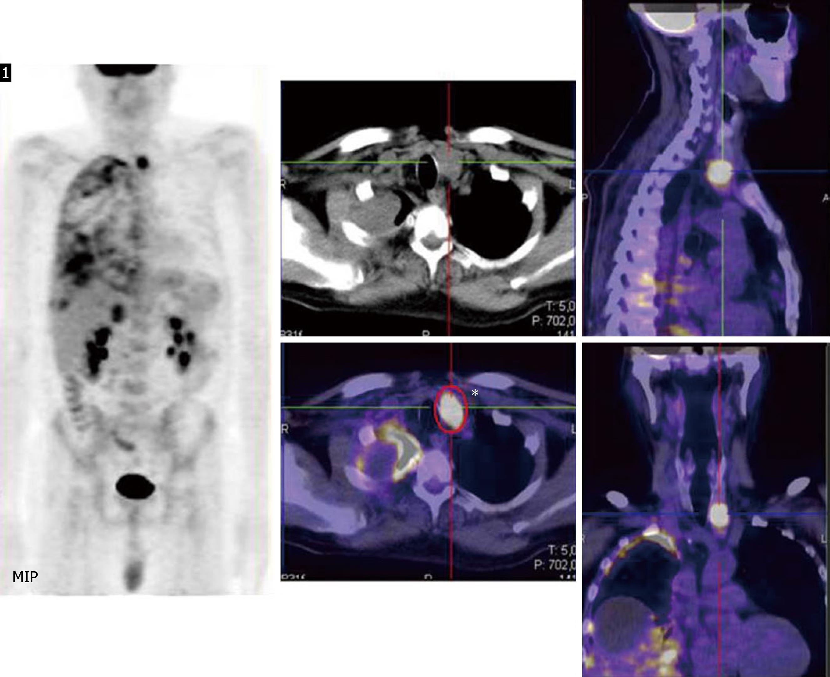
Figure 1 Staging.
Right: At Maximum intensity projection the parathyroid and pleural involvements are visible; Central and left: High 18F-Fluorodeoxyglucose-uptake is clearly displayed corresponding to the left parathyroid gland and right pleura in coronal, transverse and sagittal planes.
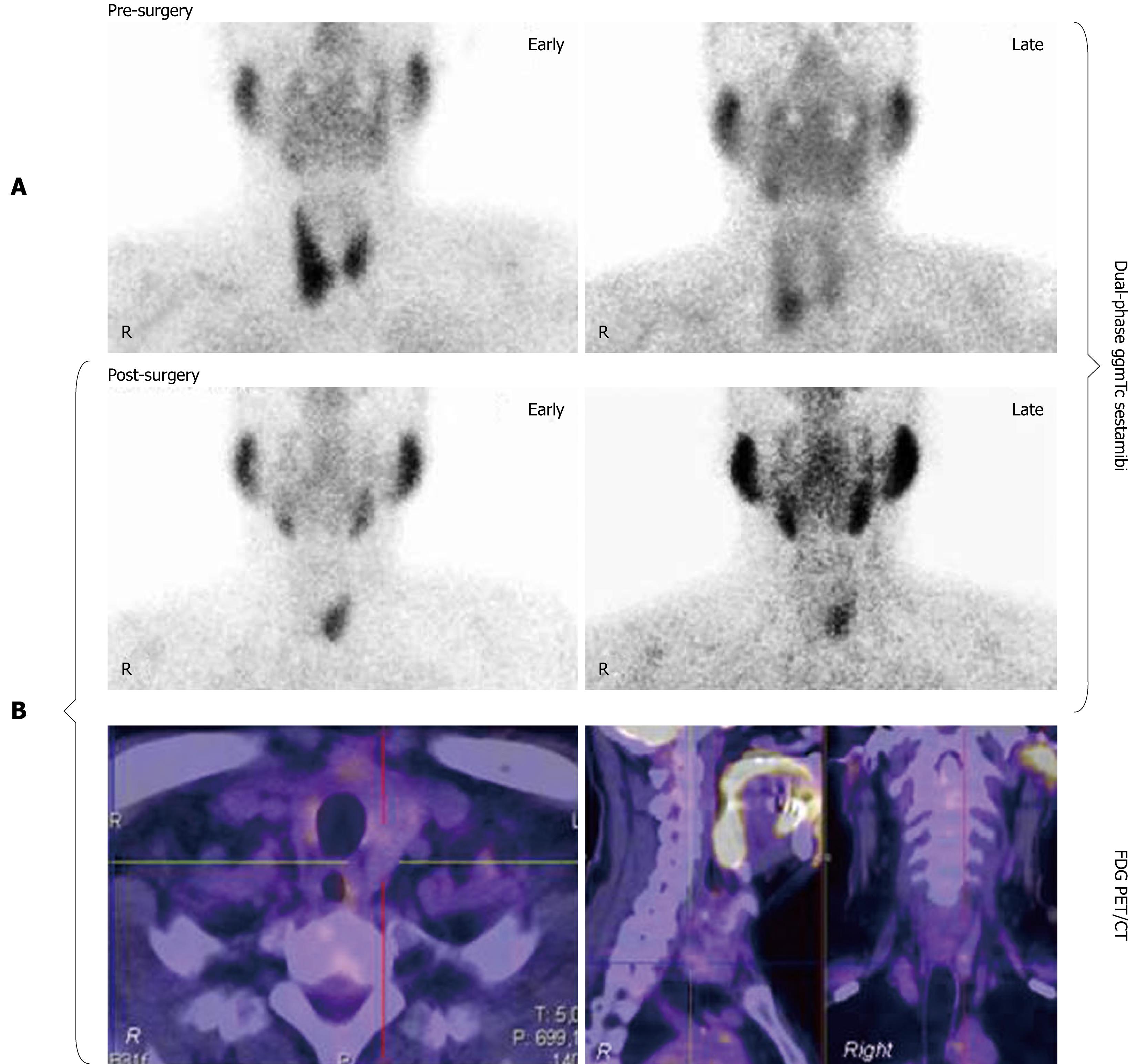
Figure 2 Restaging.
A: Pre-surgical 99mTc99 sestamibi early (15 min) and delayed (2 h) images showed the presence of high uptake in the right inferior parathyroid gland; B: Post-surgical scintigraphy scan demonstrated high tracer uptake in the left inferior parathyroid gland at early and late images, whereas no 18F-Fluorodeoxyglucose-uptake in the same gland during the positron emission tomography/computed tomography scan was reported.
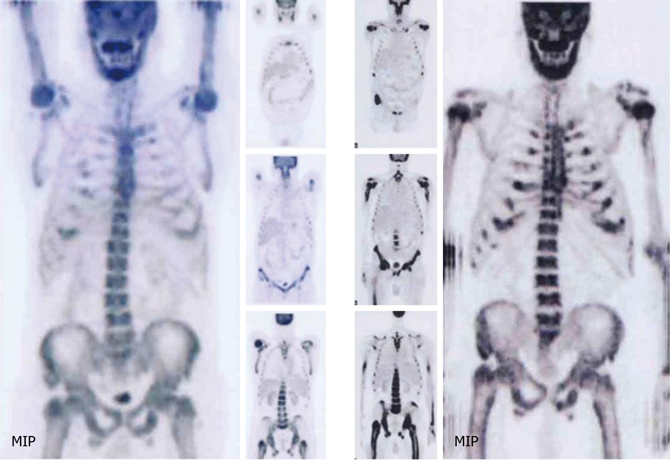
Figure 3 Progression of disease.
Right: First positron emission tomography/computed tomography (PET/CT) exam demonstrated moderate and diffuse skeletal involvement, particularly in medullar bone; Left: Second PET/CT scan, performed 5 mo after earlier PET exam, showed progression of skeletal disease.
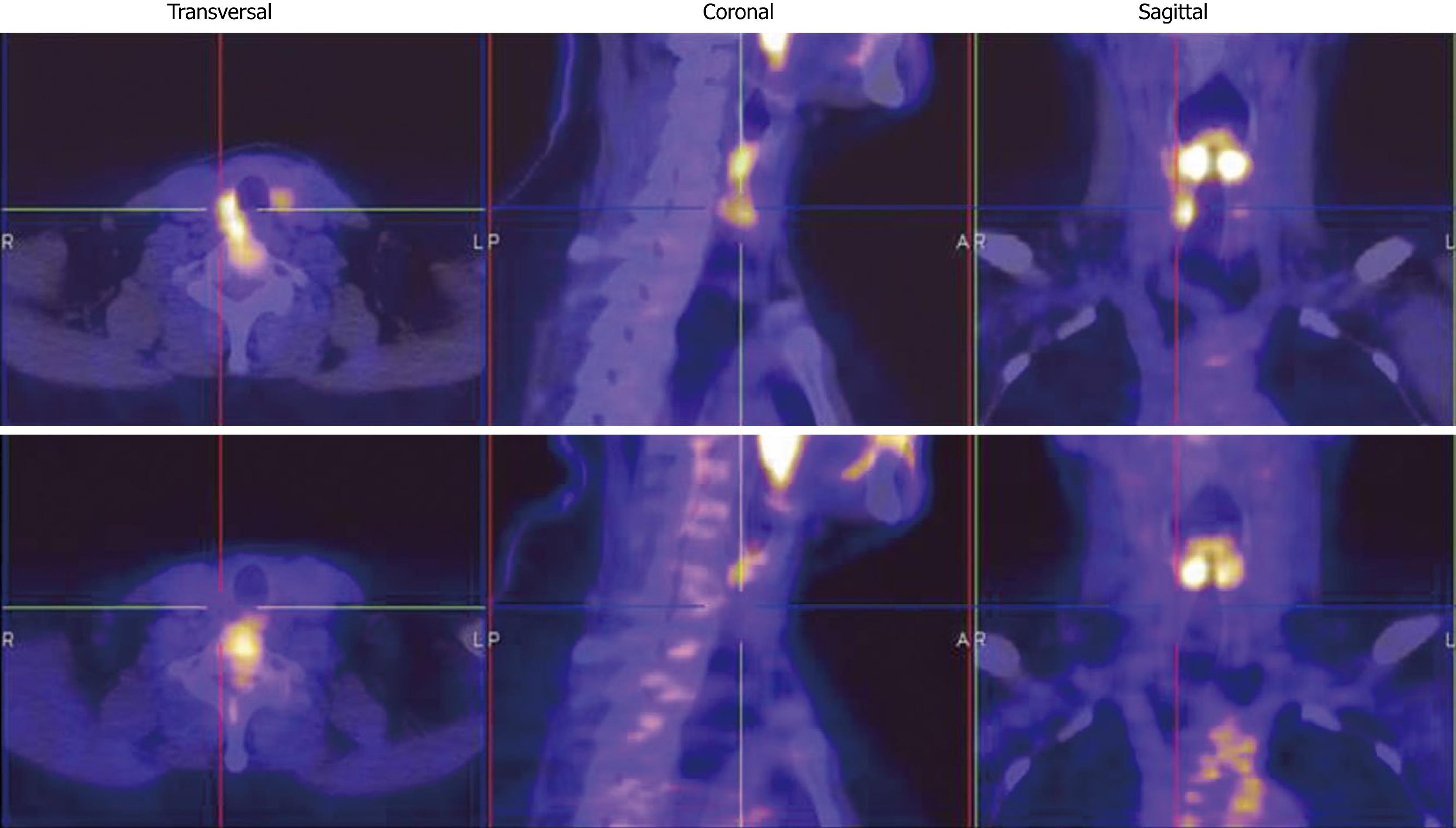
Figure 4 Post-surgical evaluation.
Up: First positron emission tomography/computed tomography (PET/CT) scan performed at least 50 d after curative surgery, demonstrated mild 18F-Fluorodeoxyglucose (FDG) uptake corresponding to the right lateral wall of the larynx; Down: Second PET/CT exam, performed 4 mo after curative surgery, demonstrated the disappearance of FDG-uptake at the same site.
- Citation: Evangelista L, Sorgato N, Torresan F, Boschin IM, Pennelli G, Saladini G, Piotto A, Rubello D, Pelizzo MR. FDG-PET/CT and parathyroid carcinoma: Review of literature and illustrative case series. World J Clin Oncol 2011; 2(10): 348-354
- URL: https://www.wjgnet.com/2218-4333/full/v2/i10/348.htm
- DOI: https://dx.doi.org/10.5306/wjco.v2.i10.348


