Copyright
©2011 Baishideng Publishing Group Co.
World J Clin Oncol. Oct 10, 2011; 2(10): 344-347
Published online Oct 10, 2011. doi: 10.5306/wjco.v2.i10.344
Published online Oct 10, 2011. doi: 10.5306/wjco.v2.i10.344
Figure 1 Coronal computed tomography of the head revealing a large destructive invasive mass involving primarily the left maxillary sinus and left ethmoidal air cells with intracranial and intraorbital extension.
Figure 2 Transverse computed tomography revealing a large destructive invasive mass involving primarily the left maxillary sinus and left ethmoidal air cells with intracranial and intraorbital extension.
Figure 3 Coronal computed tomography scan demonstrated a large enhancing soft tissue mass centered in the right ostiomeatal complex extending into the right maxillary sinus, right ethmoid air cells and right nasal cavity.
Figure 4 Status post right enucleation, right maxillectomy and hemipalatectomy, with removal of the nasal septum, excision of the right superior, middle and inferior turbinates and right ethmoidectomy.
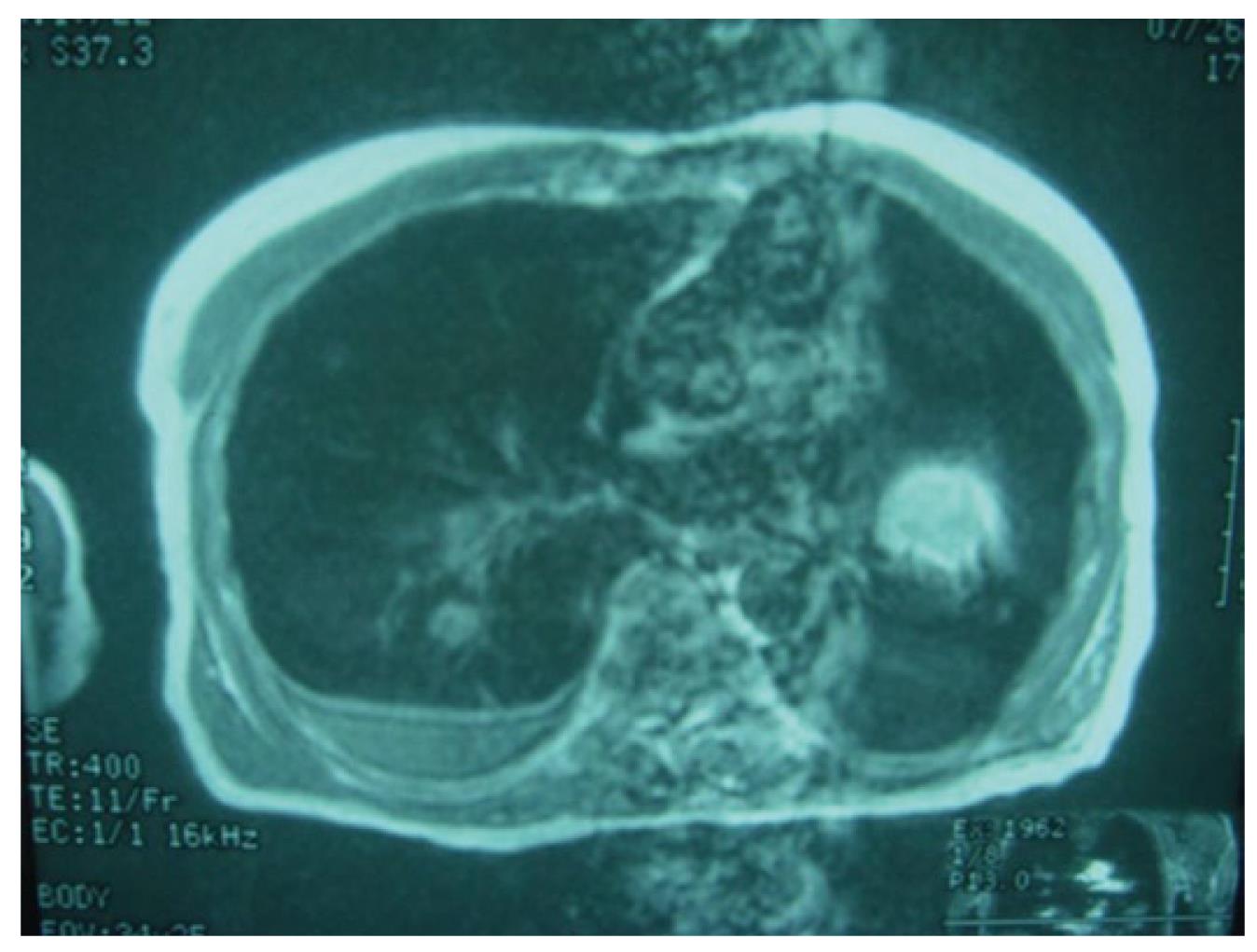
Figure 5 Transverse computed tomography of the chest demonstrated bilateral lung masses with calcifications and at least two discrete nodules suspicious for metastatic disease.
Figure 6 S100 and homatropine methylbromide (HMB45) staining positive for melanoma.
- Citation: Gasparyan A, Amiri F, Safdieh J, Reid V, Cirincione E, Shah D. Malignant mucosal melanoma of the paranasal sinuses: Two case presentations. World J Clin Oncol 2011; 2(10): 344-347
- URL: https://www.wjgnet.com/2218-4333/full/v2/i10/344.htm
- DOI: https://dx.doi.org/10.5306/wjco.v2.i10.344


