Copyright
©The Author(s) 2025.
World J Clin Oncol. Aug 24, 2025; 16(8): 109934
Published online Aug 24, 2025. doi: 10.5306/wjco.v16.i8.109934
Published online Aug 24, 2025. doi: 10.5306/wjco.v16.i8.109934
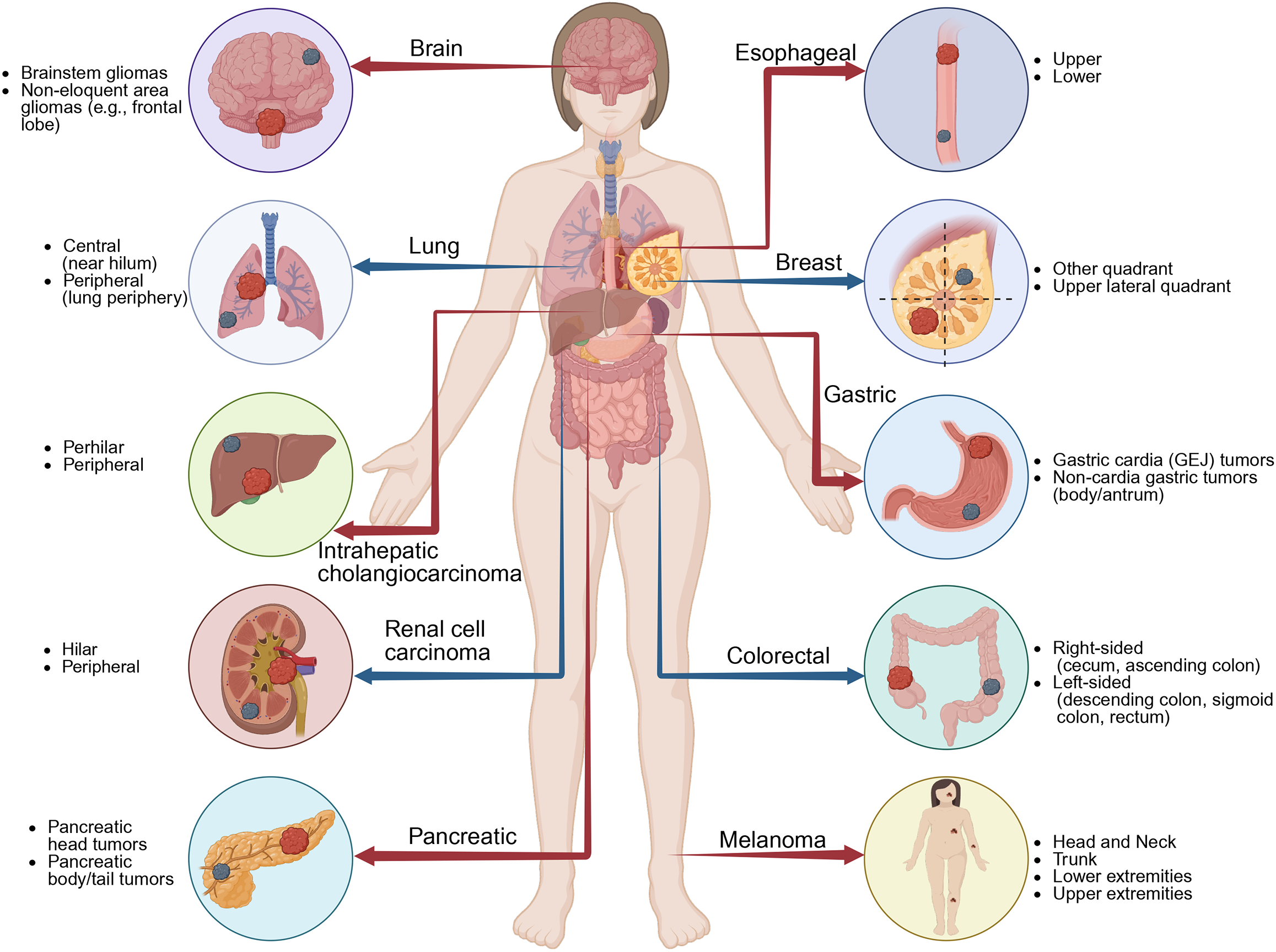
Figure 1 Tumor types where anatomical location affects prognosis.
It illustrates multiple cancer types where tumor prognosis varies with anatomical location. Examples include brain tumors (brainstem vs non-eloquent regions), lung cancer (central vs peripheral), intrahepatic cholangiocarcinoma (perihilar vs peripheral), renal cell carcinoma (hilar vs peripheral), pancreatic cancer (head vs body/tail), esophageal cancer (upper vs lower), breast cancer (upper lateral quadrant vs other quadrants), gastric cancer (cardia vs non-cardia), colorectal cancer (right-sided vs left-sided), and melanoma (head and neck, trunk, upper extremities, lower extremities). GEJ: Gastroesophageal junction.
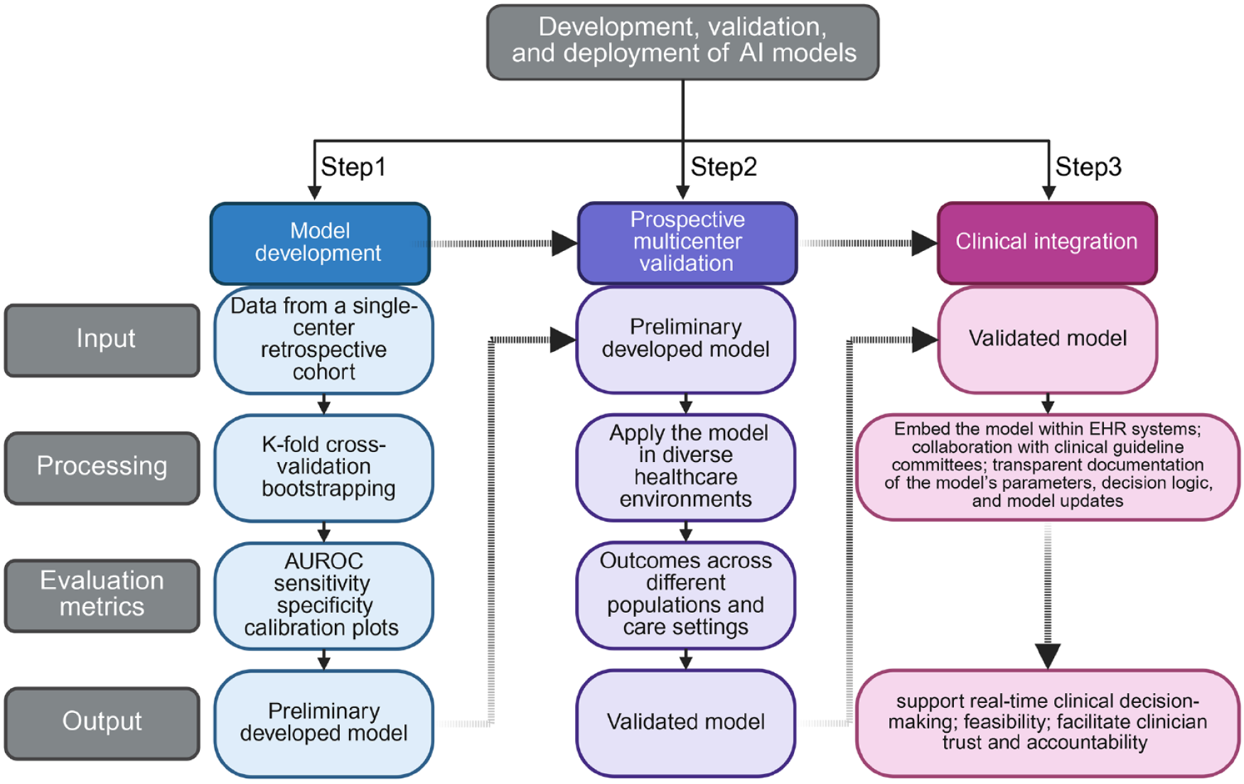
Figure 2 Standardized pipeline for development, validation, and clinical deployment of artificial intelligence models in clinical settings.
The schematic illustrates a three-step framework: (1) Model development involves training using single-center retrospective data, with internal validation via k-fold cross-validation and bootstrapping, and performance assessed by area under the receiver operating characteristic curve, sensitivity, specificity, and calibration plots; (2) Prospective multicenter validation applies the preliminary model across heterogeneous clinical settings to evaluate generalizability and establish external validity. This is crucial to ensure the model performs equally good across external populations; and (3) Clinical integration incorporates the validated model into Electronic Health Record systems, with guideline committee collaboration and transparent documentation of parameters, decision logic, and updates to enable real-time clinical use and support provider confidence. AUROC: Area under the receiver operating characteristic.
- Citation: Wang C, Chen MY, Wang YG, Shi M. Integrating tumor location into artificial intelligence-based prognostic models in cancer. World J Clin Oncol 2025; 16(8): 109934
- URL: https://www.wjgnet.com/2218-4333/full/v16/i8/109934.htm
- DOI: https://dx.doi.org/10.5306/wjco.v16.i8.109934


