Copyright
©The Author(s) 2025.
World J Clin Oncol. Aug 24, 2025; 16(8): 109088
Published online Aug 24, 2025. doi: 10.5306/wjco.v16.i8.109088
Published online Aug 24, 2025. doi: 10.5306/wjco.v16.i8.109088
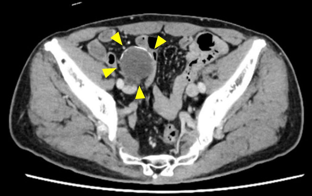
Figure 1 Abdominal contrast-enhanced computed tomography scan (axial view).
A 38-mm cystic mass was observed within the appendix. It features a non-enhanced hypodense area and surrounding curvilinear calcifications (yellow arrowheads).
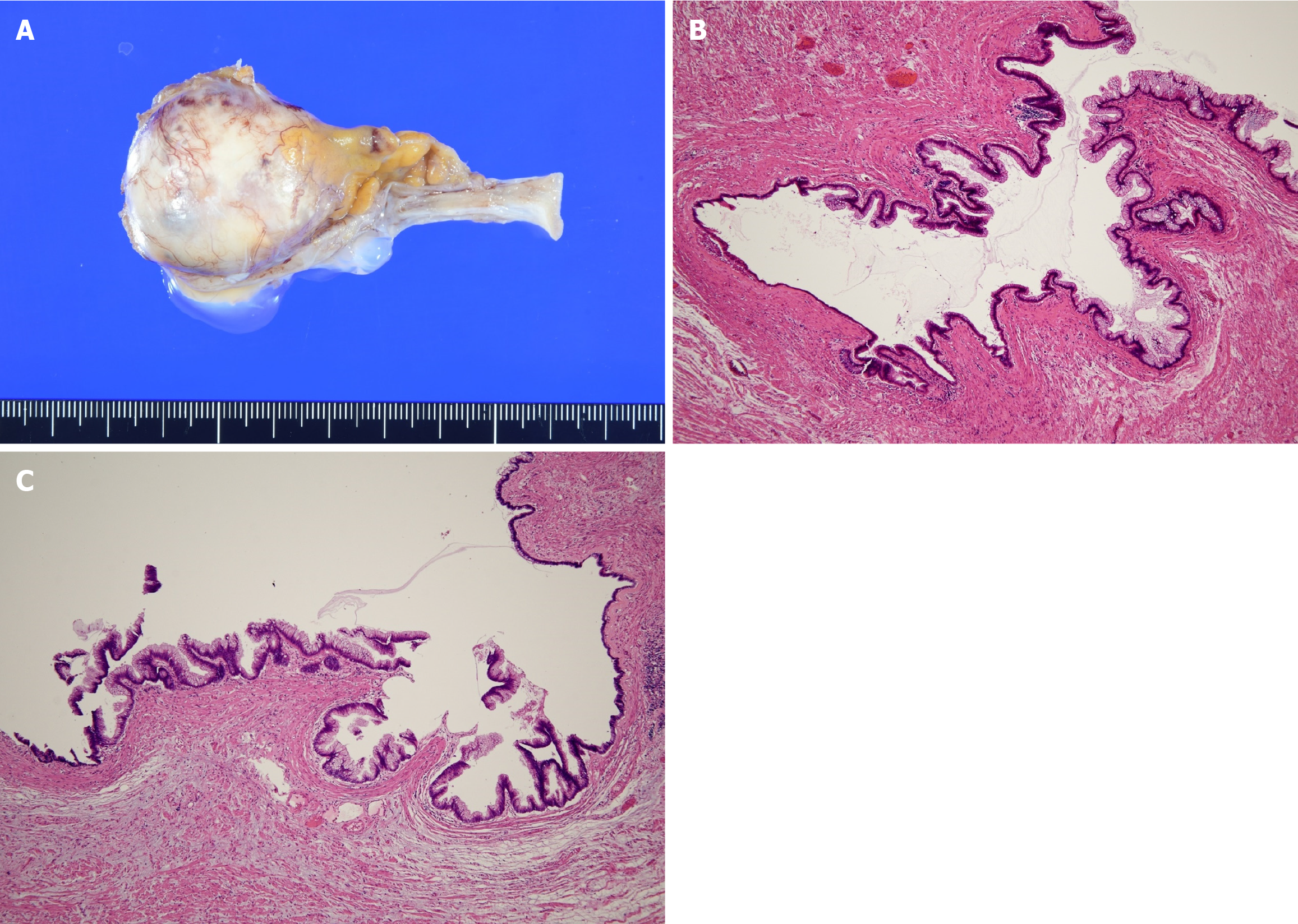
Figure 2 Macroscopic and microscopic findings.
A: Macroscopic view of a low-grade appendiceal mucinous neoplasm (LAMN), showing cystic dilation of the appendix filled with mucinous content; B: Microscopic view of a LAMN with hematoxylin & eosin staining (× 40 magnification), showing a mucosal surface lined by a single layer of mucin-producing columnar epithelium without high-grade cytologic atypia; C: Additional microscopic view of a LAMN with hematoxylin & eosin staining (× 40 magnification), highlighting features similar to panel B.
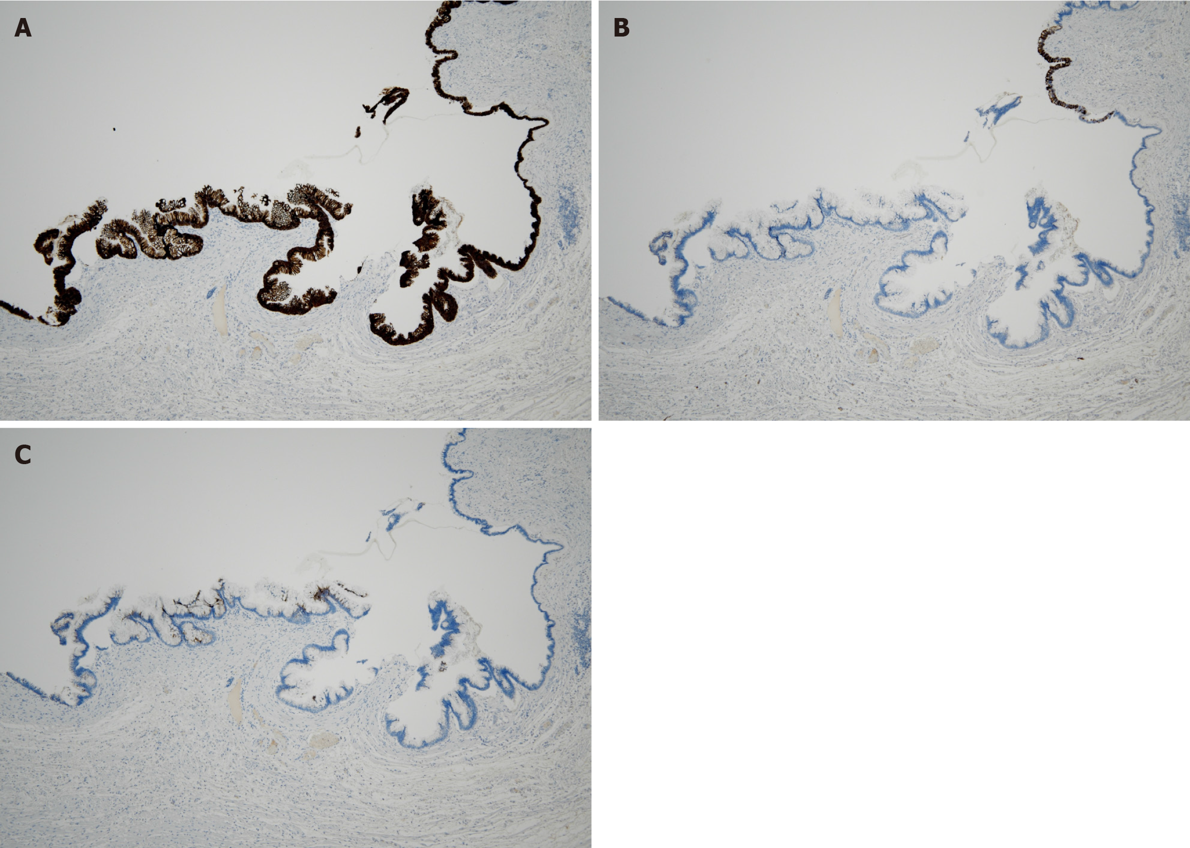
Figure 3 Immunohistochemical findings.
A: Immunohistochemical staining for cytokeratin 20, showing diffuse positivity on the mucosal surface layer (× 40 magnification); B: Immunohistochemical staining of cytokeratin 7, showing partial positivity on the mucosal surface layer (× 40 magnification); C: Immunohistochemical staining for mucin 5AC, showing partial positivity on the mucosal surface layer (× 40 magnification).
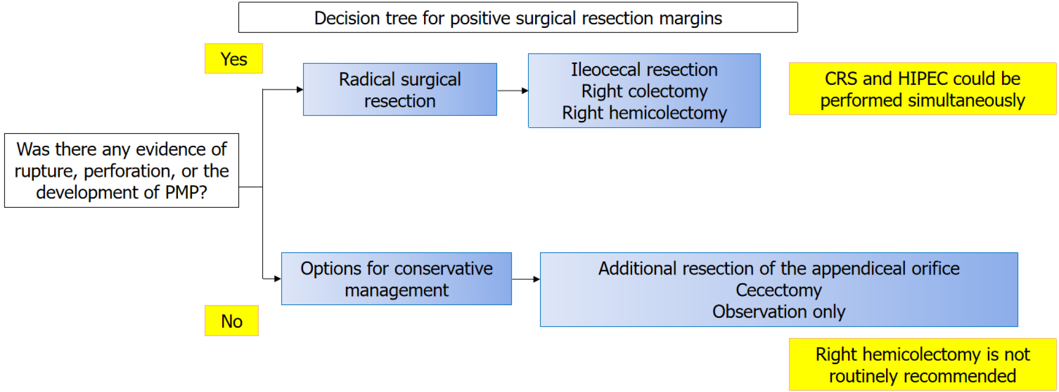
Figure 4 Decision tree for positive surgical resection margins.
CRS: Cytoreductive surgery; HIPEC: Hyperthermic intraperitoneal chemotherapy; PMP: Pseudomyxoma peritonei.
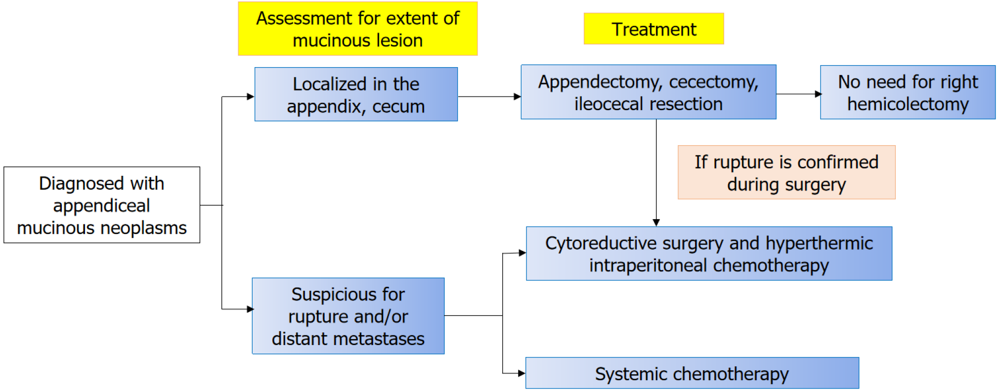
Figure 5
Proposed algorithm for the treatment strategy of appendiceal mucinous neoplasms.
- Citation: Mitamura A, Tsujinaka S, Fujishima F, Sawada K, Hikage M, Miura T, Kitamura Y, Hatsuzawa Y, Nakano T, Shibata C. Appendiceal mucinous neoplasms: Optimizing treatment strategies based on clinical, histological, and molecular features. World J Clin Oncol 2025; 16(8): 109088
- URL: https://www.wjgnet.com/2218-4333/full/v16/i8/109088.htm
- DOI: https://dx.doi.org/10.5306/wjco.v16.i8.109088


