Copyright
©The Author(s) 2025.
World J Clin Oncol. Apr 24, 2025; 16(4): 104413
Published online Apr 24, 2025. doi: 10.5306/wjco.v16.i4.104413
Published online Apr 24, 2025. doi: 10.5306/wjco.v16.i4.104413
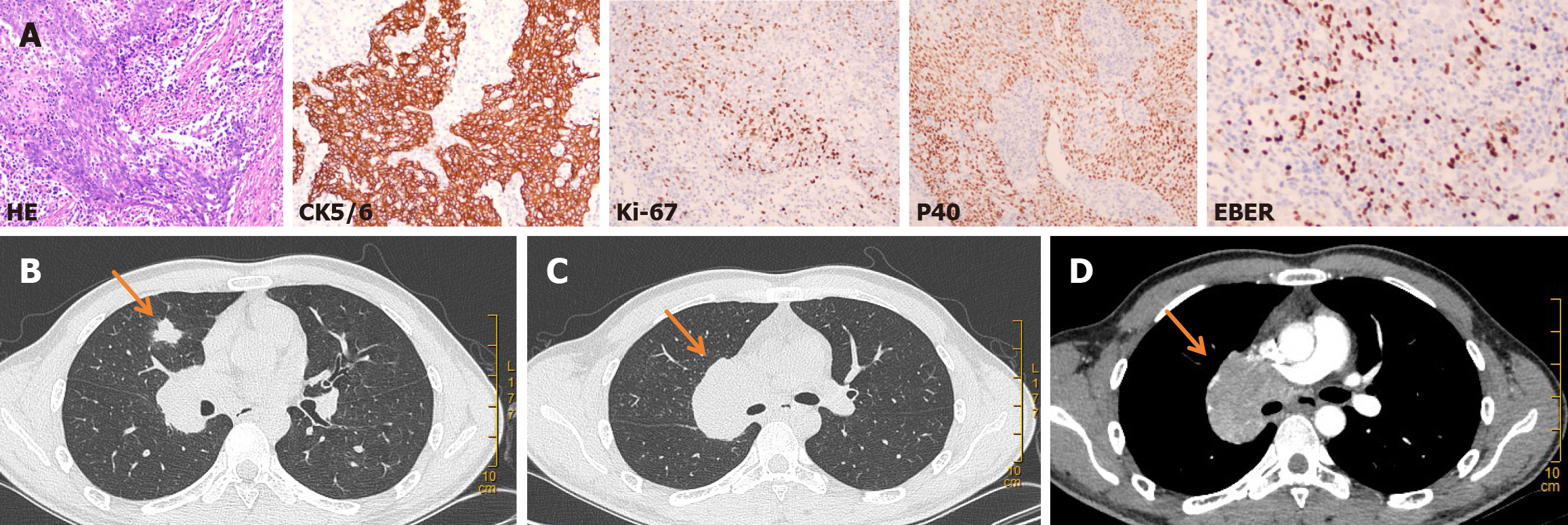
Figure 1 Right pulmonary lymphoepithelioma-like carcinoma.
A: Histopathology of the middle lobe of the right lung (hematoxylin and eosin staining; magnification × 200): Pulmonary lymphoepithelioma-like carcinoma, tumor cells are arranged in a nest-like pattern, accompanied by massive lymphocyte infiltration. Immunohistochemistry: Cytokeratin 5/6 (+), Ki67 (+), P40 (+); in situ hybridization: Epstein-Barr encoding region (+); B: A mass in the middle lobe of the right lung (approximately 1.6 cm × 1.7 cm); C and D: Lymph node metastasis in the mediastinum and right hilum of the lung (the largest lymph node, approximately 5.4 cm × 4.2 cm, was located in the right hilum). CK5/6: Cytokeratin 5/6; HE: Hematoxylin and eosin; EBER: Epstein-Barr encoding region.
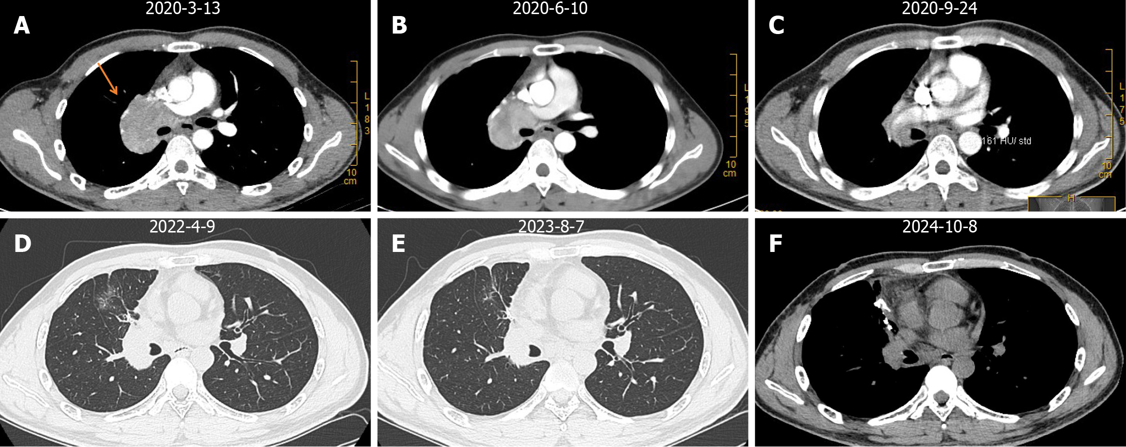
Figure 2 Postoperative follow-up status of lymph node metastasis in the mediastinum and right hilum of the lung.
A: Before surgical treatment; B: After 4 cycles of toripalimab treatment; C: After 10 cycles of toripalimab treatment; D-F: Follow-up imaging after surgery. The therapeutic effect was evaluated as stable disease.
- Citation: Huang FL, Luo M, He ZM, Shen YQ, Liu GD. Diagnosis and treatment of pulmonary lymphoepithelioma-like carcinoma: A case report. World J Clin Oncol 2025; 16(4): 104413
- URL: https://www.wjgnet.com/2218-4333/full/v16/i4/104413.htm
- DOI: https://dx.doi.org/10.5306/wjco.v16.i4.104413


