Copyright
©The Author(s) 2025.
World J Clin Oncol. Apr 24, 2025; 16(4): 103651
Published online Apr 24, 2025. doi: 10.5306/wjco.v16.i4.103651
Published online Apr 24, 2025. doi: 10.5306/wjco.v16.i4.103651
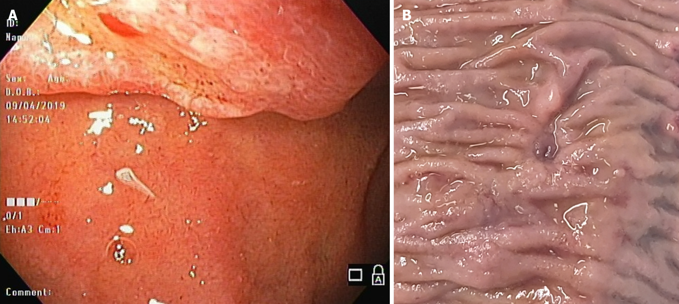
Figure 1 Ampullary adenocarcinoma.
A: Endoscopic image at diagnosis (courtesy of Dr. Carlos León); B: Macroscopic image of surgical specimen after adjuvant treatment.
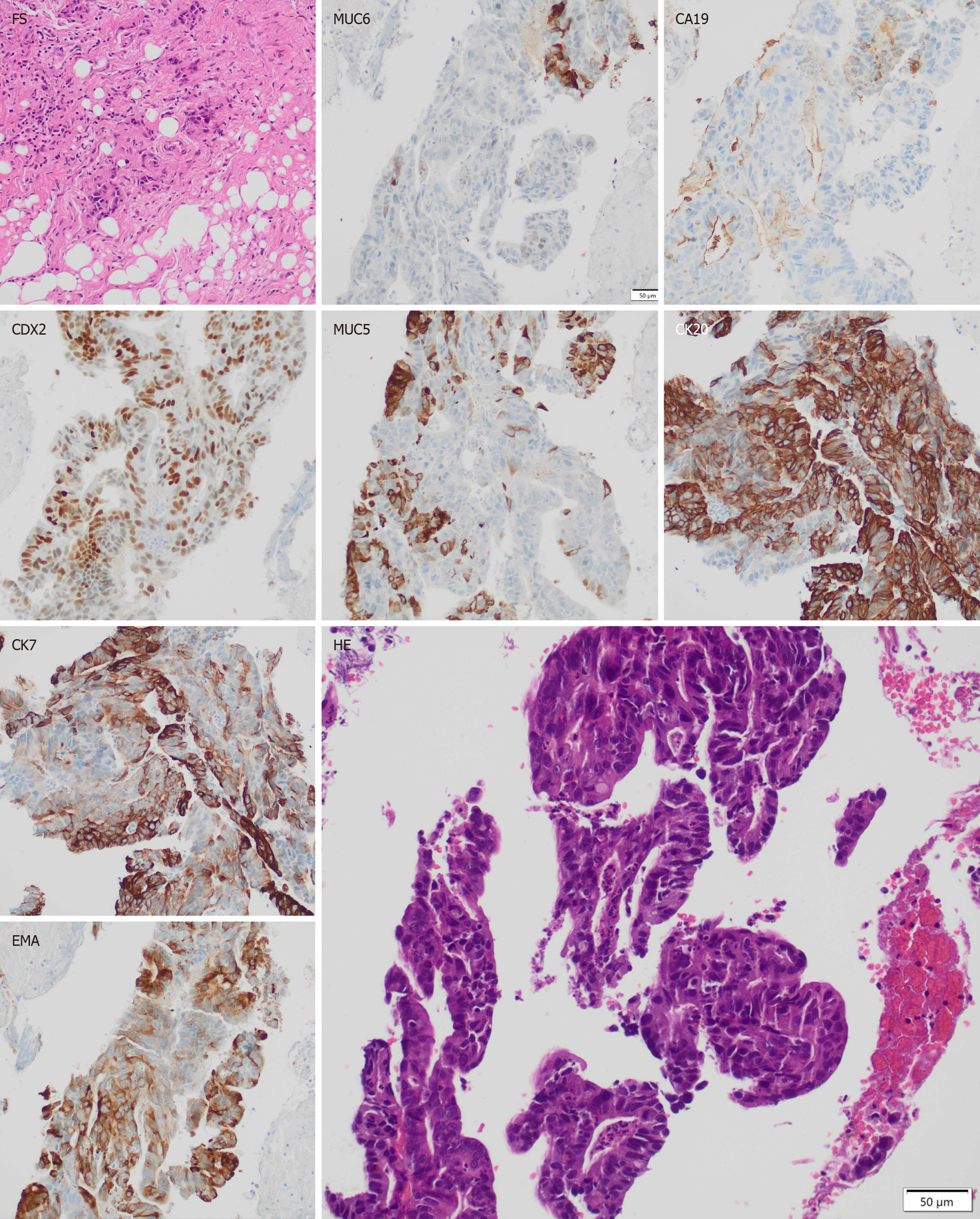
Figure 2 Histology.
An intraoperative frozen section was obtained from peritoneal mesentery. It evidences a carcinoma. The other samples were taken from the primary tumor and analysed in a delayed study. The hematoxylin-eosine stain shows an ampullary adenocarcinoma. The immunohistochemical study evidences an ampullary adenocarcinoma with mixed profile. Intense positivity for caudal-related homeobox transcription factor 2, cytokeratin 20 are indicative of intestinal type differentiation, while positivity for carbohydrate antigen 19-9, epithelial membrane antigen and mucin 6 demonstrate minor biliopancreatic differentiation. Mucin 5AC and cytokeratin 7 markers can be seen in both histologic types. (All the histological images were taken using a 20 × objective). FS: Frozen section; HE: Hematoxylin-eosine stain; CDX2: Caudal-related homeobox transcription factor 2; CA 19-9: Carbohydrate antigen 19-9; MUC6: Mucin 6; MUC5AC: Mucin 5AC; CK7: Cytokeratin 7; EMA: Epithelial membrane antigen; CK20: Cytokeratin 20.
- Citation: Núñez JC, Rivera MT, Stevens MA. Adenocarcinoma of the duodenal papilla with synchronous peritoneal metastases-5 years of overall survival: A case report. World J Clin Oncol 2025; 16(4): 103651
- URL: https://www.wjgnet.com/2218-4333/full/v16/i4/103651.htm
- DOI: https://dx.doi.org/10.5306/wjco.v16.i4.103651


