Copyright
©The Author(s) 2025.
World J Clin Oncol. Apr 24, 2025; 16(4): 102297
Published online Apr 24, 2025. doi: 10.5306/wjco.v16.i4.102297
Published online Apr 24, 2025. doi: 10.5306/wjco.v16.i4.102297
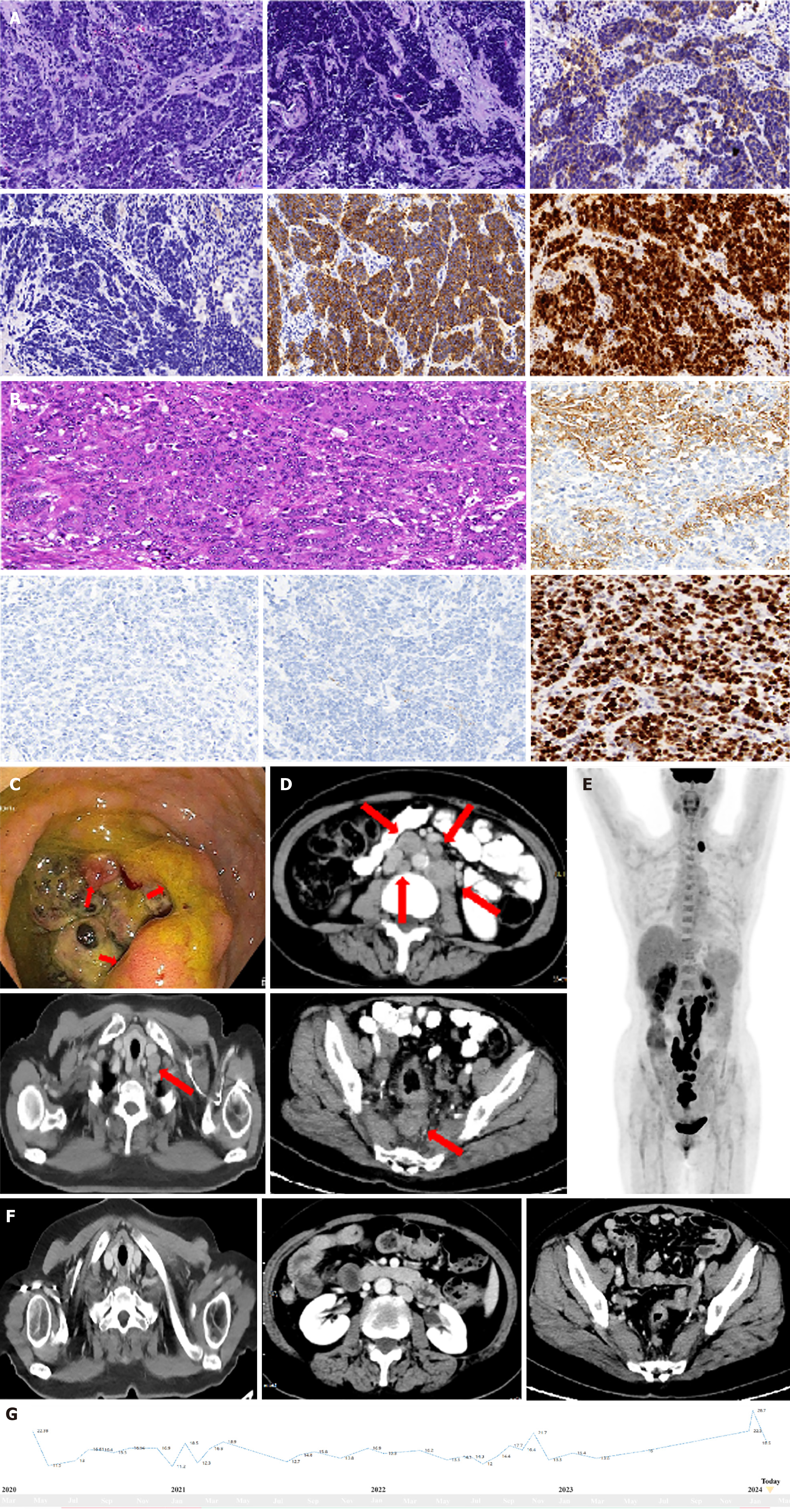
Figure 1 Imaging examination, laboratory examination for cases.
A: HE staining revealed poorly differentiated atypical cells with mixed large and small cells; the scale bar at the bottom right represents 1 cm. Immunohistochemistry of Syn, CgA, CD56 and ki-67; B: Pathological assessment of surgical tissue slides: HE staining revealed poorly differentiated ulcerous mixed neuroendocrine/neuroendocrine carcinoma (MiNEC), with a ratio of 30% poorly differentiated adenocarcinoma to MiNEC; C: Colonoscopy revealed a circumferential rectal neoplasm with an irregular shape; D: Computed tomography (CT) at diagnosis revealed irregular thickening of the rectal wall with uneven enhancement and multiple enlarged nodes in the pelvic cavity, abdomen, and left neck; E: PET/CT at diagnosis revealed hypermetabolic nodes in the pelvic cavity, abdomen, and left neck; F: CTs after 11 cycles of camrelizumab and surufatinib revealed partial remission; the sizes of both the primary rectal lesion and the lymph nodes had decreased (more than 30%); G: Dynamics of NSE during the treatment process.
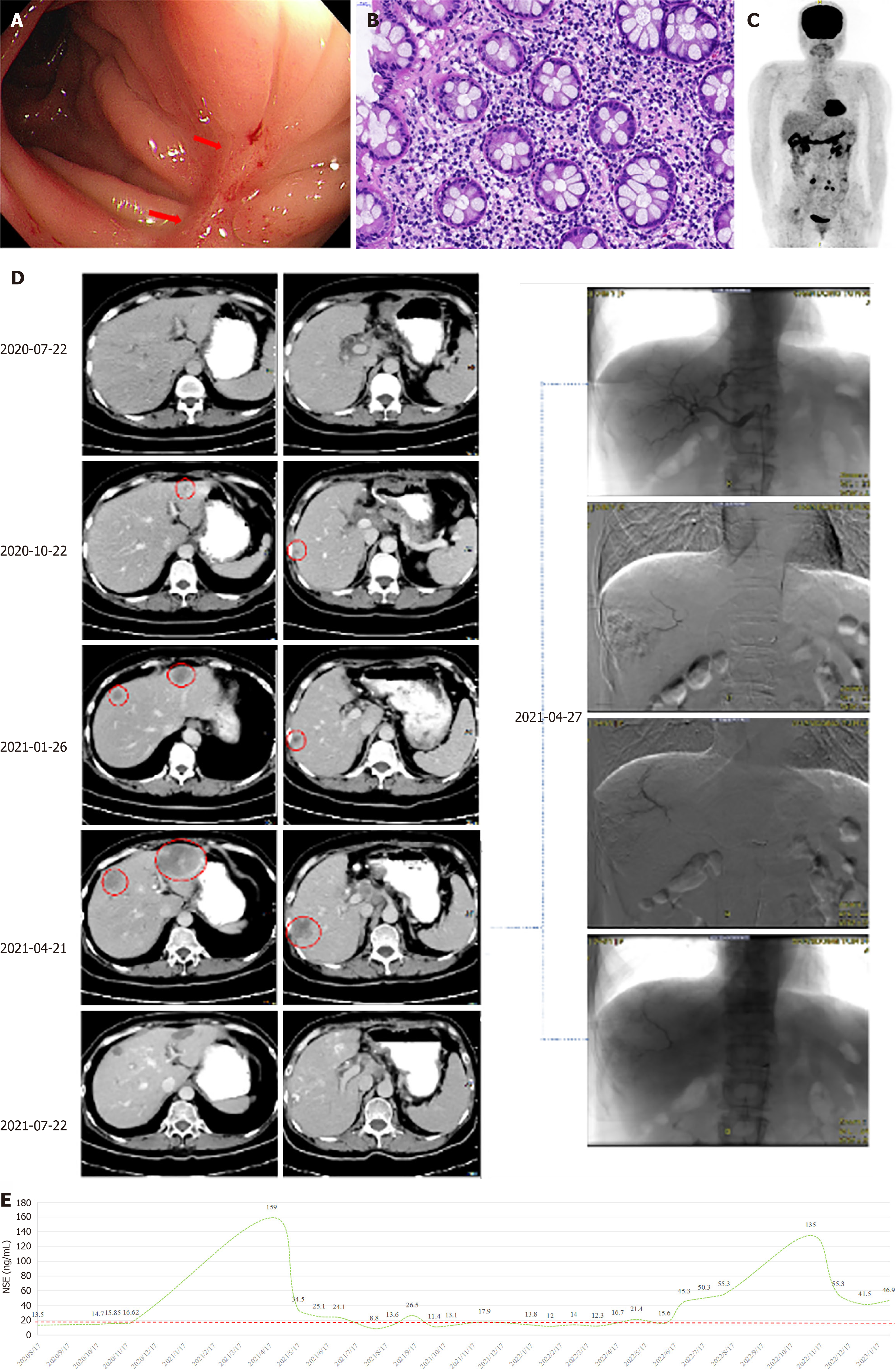
Figure 2 Imaging and laboratory tests of these cases and the course of TACE treatment of the patient in case 2.
A: Colonoscopy revealed scar-like changes in the original rectal lesion with distorted surrounding mucosa; B: Microscopy of the biopsy sample revealed chronic inflammation with lymphocyte infiltration; C: PET/CT revealed no hypermetabolic uptake in the rectum or left neck, with several residual lymph nodes in the abdomen. Computed tomography (CT) at diagnosis revealed irregular thickening of the rectal wall with uneven enhancement and multiple enlarged nodes in the pelvic cavity, abdomen, and left neck; D: CT images at 1 month after surgery, 4 cycles (PD) of etoposide + carboplatin, 3 cycles (PD) of irinotecan + raltitrexed, and 3 cycles (PD) of xelox + bevacizumab. Sintilimab + surufatinib (ICI-TKI) continuous treatment (TACE of hepatic metastasis 2021-04-27); E: Dynamics of NSE during the treatment process.
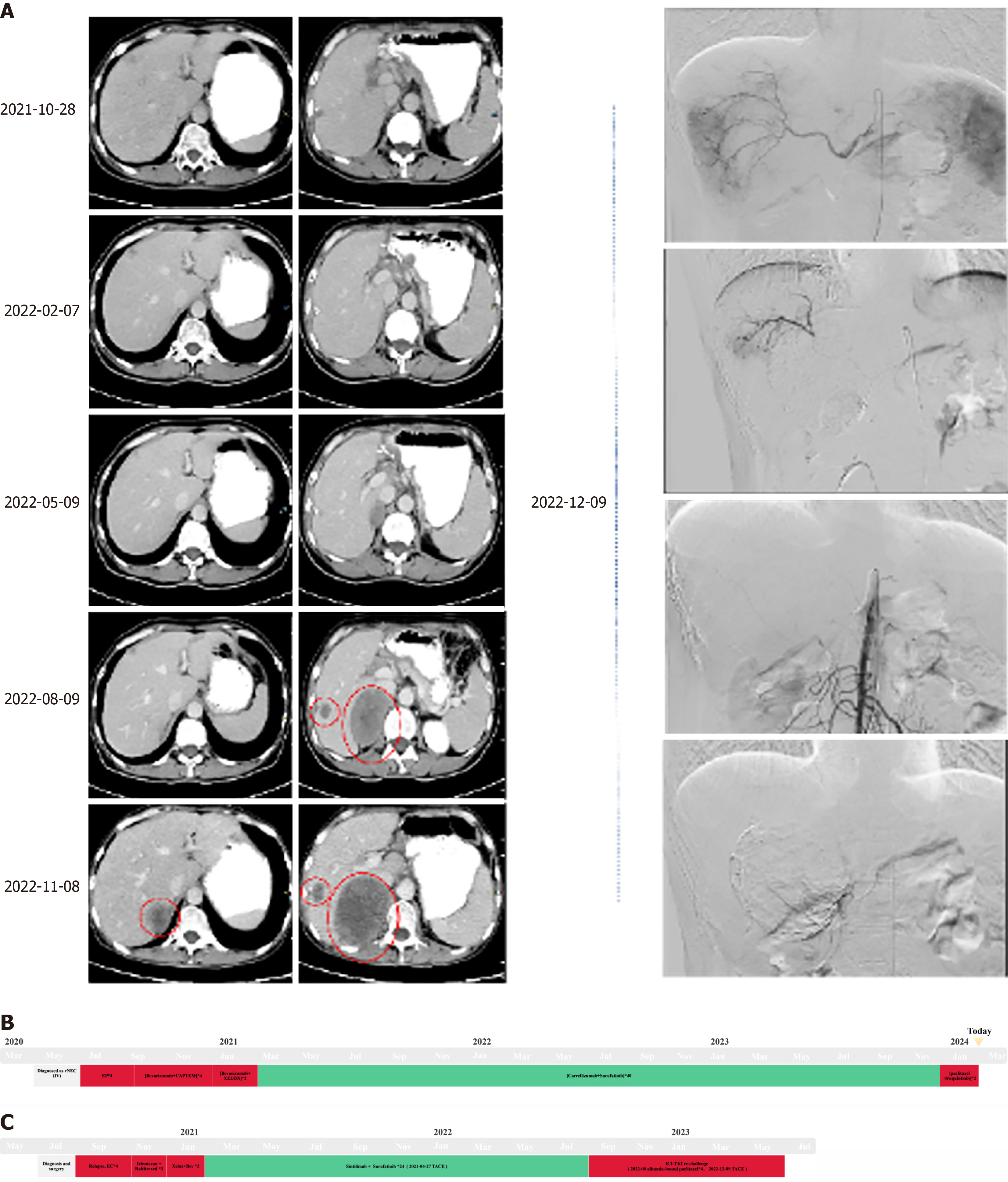
Figure 3 The treatment history of arterial embolism in patient 2 and the treatment plan for these two patients.
A: ICI-TKI continuous treatment, left adrenal gland and hepatic metastasis chemoembolization, and X cycles of ICI-TKI rechallenge; B: Timeline of the diagnosis and treatment process for case 1; C: Timeline of the diagnosis and treatment process for case 2.
- Citation: Gao LL, Gao DN, Yuan HT, Chen WQ, Yang J, Peng JQ. Combining anti-PD-1 antibodies with surufatinib for gastrointestinal neuroendocrine carcinoma: Two cases report and review of literature. World J Clin Oncol 2025; 16(4): 102297
- URL: https://www.wjgnet.com/2218-4333/full/v16/i4/102297.htm
- DOI: https://dx.doi.org/10.5306/wjco.v16.i4.102297


